

Modulation of Nitric Oxide Synthases by Oxidized LDLs: Role in Vascular Inflammation and Atherosclerosis Development
- PMID: 31277498
- PMCID: PMC6651385
- DOI: 10.3390/ijms20133294
Modulation of Nitric Oxide Synthases by Oxidized LDLs: Role in Vascular Inflammation and Atherosclerosis Development
Abstract
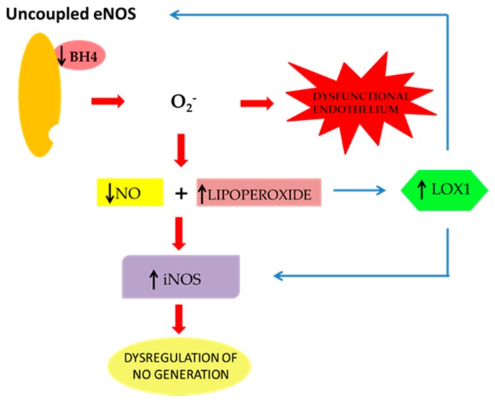
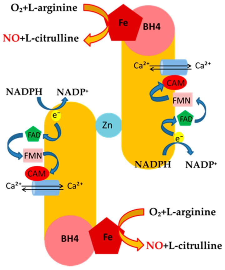
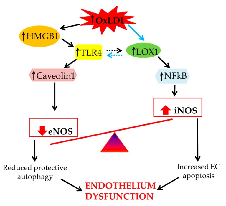
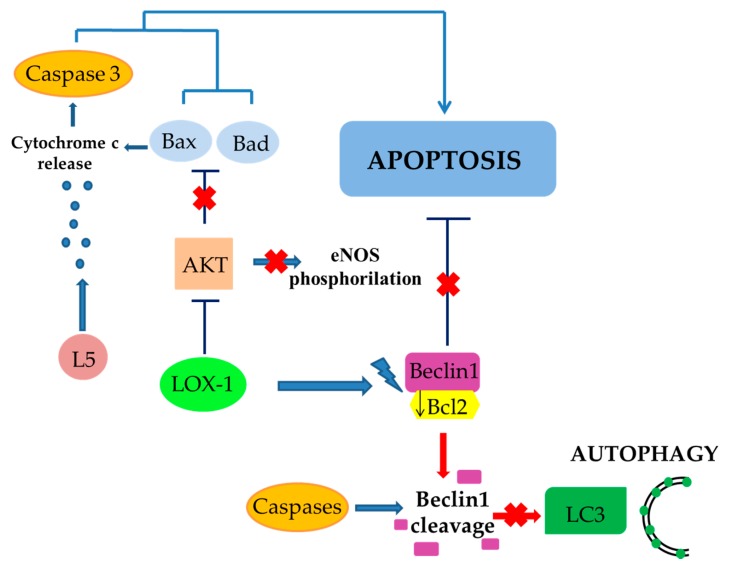
The maintenance of physiological levels of nitric oxide (NO) produced by eNOS represents a key element for vascular endothelial homeostasis. On the other hand, NO overproduction, due to the activation of iNOS under different stress conditions, leads to endothelial dysfunction and, in the late stages, to the development of atherosclerosis. Oxidized LDLs (oxLDLs) represent the major candidates to trigger biomolecular processes accompanying endothelial dysfunction and vascular inflammation leading to atherosclerosis, though the pathophysiological mechanism still remains to be elucidated. Here, we summarize recent evidence suggesting that oxLDLs produce significant impairment in the modulation of the eNOS/iNOS machinery, downregulating eNOS via the HMGB1-TLR4-Caveolin-1 pathway. On the other hand, increased oxLDLs lead to sustained activation of the scavenger receptor LOX-1 and, subsequently, to NFkB activation, which, in turn, increases iNOS, leading to EC oxidative stress. Finally, these events are associated with reduced protective autophagic response and accelerated apoptotic EC death, which activates atherosclerotic development. Taken together, this information sheds new light on the pathophysiological mechanisms of oxLDL-related impairment of EC functionality and opens new perspectives in atherothrombosis prevention.
Keywords: constitutive NO synthase cNOS; endothelial dysfunction; inducible NO synthase (iNOS); oxidized LDLs.
Conflict of interest statement
The authors declare no conflict of interest.
Figures
References
-
- Maiuolo J., Gliozzi M., Musolino V., Scicchitano M., Carresi C., Scarano F., Bosco F., Nucera S., Ruga S., Zito M.C., et al. The “Frail” Brain Blood Barrier in Neurodegenerative Diseases: Role of Early Disruption of Endothelial Cell-to-Cell Connections. Int. J. Mol. Sci. 2018;19:2693. doi: 10.3390/ijms19092693. - DOI - PMC - PubMed
-
- Lanuti P., Rotta G., Almici C., Avvisati G., Budillon A., Doretto P., Malara N., Marini M., Neva A., Simeone P., et al. Endothelial progenitor cells, defined by the simultaneous surface expression of VEGFR2 and CD133, are not detectable in healthy peripheral and cord blood. Cytom. A. 2016;89:259–270. doi: 10.1002/cyto.a.22730. - DOI - PubMed
-
- Malara N.M., Trunzo V., Musolino G., Aprigliano S., Rotta G., Macrina L., Limongi T., Gratteri S., di Fabrizio E., Renzulli A., et al. Soluble CD54 induces human endothelial cells ex vivo expansion useful for cardiovascular regeneration and tissue engineering application. Int. J. Cardiol. Heart Vasc. 2015;6:48–53. doi: 10.1016/j.ijcha.2015.01.004. - DOI - PMC - PubMed
Publication types
MeSH terms
Substances
Grants and funding
LinkOut - more resources
Full Text Sources
Other Literature Sources
Medical
