. 2019 Jul 5;14(1):165.
doi: 10.1186/s13023-019-1147-9.

Kaposiform lymphangiomatosis and kaposiform hemangioendothelioma: similarities and differences

Affiliations

Kaposiform lymphangiomatosis and kaposiform hemangioendothelioma: similarities and differences

Yi Ji et al. Orphanet J Rare Dis. .

Abstract

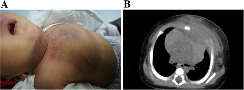
Background: Kaposiform lymphangiomatosis (KLA) and kaposiform hemangioendothelioma (KHE) are rare and aggressive vascular disorders. The aim of this study was to examine the clinical features and prognosis of KLA and KHE involving the thorax.

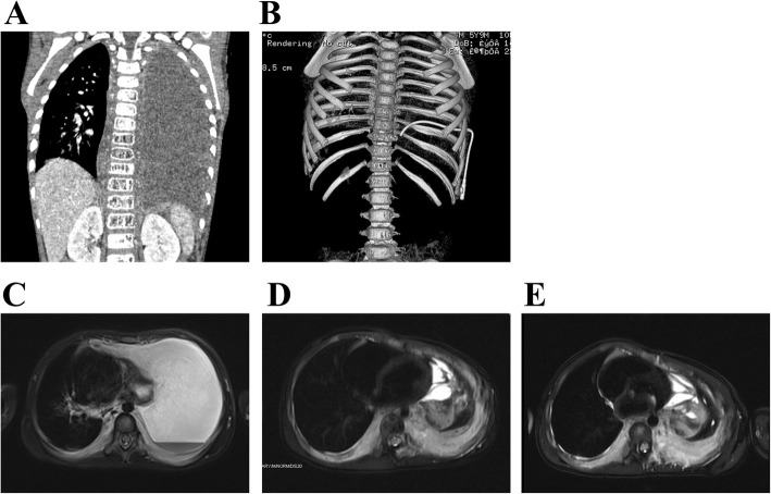
Methods: The clinical features, imaging and pathological findings, treatments and outcome were retrospectively reviewed for 6 patients with KLA and 7 patients with KHE involving the thorax.

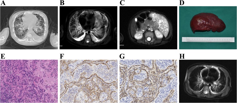
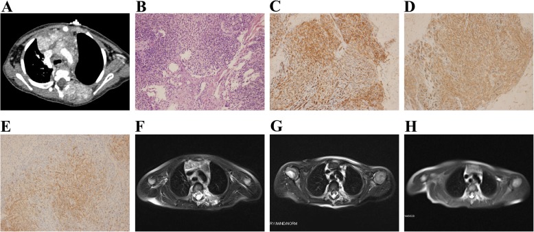
Results: The mean ages at the time of the presentation of signs/symptoms were 26.7 months and 4.1 months for KLA and KHE, respectively. Respiratory symptoms, pericardial and pleural effusion, thrombocytopenia and coagulopathy were common in both KLA and KHE. Diffuse lesions involving the lung and extrathoracic sites were observed in KLA but not in KHE. Histopathologically, all lesions had spindled tumor cells, which were immunoreactive for CD31 and D2-40. In KLA, the spindle cells were distributed in sparse and poorly marginated clusters, whereas the spindle cells formed more defined and confluent vascularized nodules in KHE. Unlike the refractory behavior of KLA, the majority of patients with KHE responded to medical treatments with regression of the lesion and normalization of the hematologic parameters.

Conclusions: The presenting and histological characteristics of KLA can overlap with those of KHE. The presence of diffuse vascular lesions in the mediastinum and lung with refractory thrombocytopenia and coagulopathy should suggest the diagnosis of KLA. Given the rarity and high morbidity and mortality of these disorders, the diagnostic process and therapeutic approach should include a multidisciplinary team review and consensus.

Keywords: Coagulopathy; Kaposiform hemangioendothelioma; Kaposiform lymphangiomatosis; Thorax; Thrombocytopenia.


Conflict of interest statement

The authors declare that they have no competing interests, either financial or non-financial, that could be perceived as prejudicing the impartiality of the research reported.

Figures

Fig. 1
Fig. 1
Radiologic findings and pathological features in KLA (Patient #3). a Unenhanced CT scan of the chest demonstrates thickening of the interlobular septa. Axial T2-weighted thoracic MRI shows pericardial effusion and high-signal abnormality in the mediastinum that extends along the bronchovascular bundles (b). Axial T2-weighted MRI of the abdomen shows multiple high-signal splenic lesions (c). Macroscopic view of the spleen excised from the patient (d). Multiple, small, reddish blue nodules are noted on the surface of the spleen. Hematoxylin-eosin staining shows dilated lymphatic channels and dispersed small irregular cellular clusters within the splenic parenchyma. The clusters are comprised of spindle-like cells without distinct lumens e. The spindle-like cells are immunopositive for CD31 (F) and D2–40 g. 6 months after splenectomy, MRI revealed a significant involution of the lesion h
Fig. 2
Fig. 2
Treatment response in patient with KLA (Patient #1). a At the initial presentation of respiratory distress, coronal enhanced CT scan revealed massive pleural effusion almost entirely filling the left hemithorax with complete atelectasis of the left lung and mediastinal shift to the right. The lesions involved the vertebral bodies of T8-T12 and multiple ribs (b). Horizontal T2-weighted MRI revealed pleural thickening and effusion, heterogeneously increased T2 signal intensity along the right tracheobronchial tree, and infiltrative, hyperintense, posterior mediastinal soft tissue masses (c). The patient was treated with vincristine for 12 months. However, the response was suboptimal. The lesion became progressively static in size (d). Treatment with sirolimus and prednisolone was then initiated. After 8 weeks of combination treatment with sirolimus and prednisolone, followed by 10 months of sirolimus monotherapy, the lesions became more prominent (e)
Fig. 3
Fig. 3
KHE with cutaneous involvement. a The photograph shows an indurated and purpuric mass in the anterior neck, chest and abdominal wall (Patient #13). Ecchymosis was evident with telangiectasia. b An unenhanced CT scan revealed an extensive soft-tissue mass with ill-defined margins throughout the anterior thoracic musculature
Fig. 4
Fig. 4
Radiologic findings, pathological features and treatment response in a patient with KHE (Patient #8). a An enhanced CT scan shows an enhanced soft-tissue mass extending from the posterior thoracic musculature to the vertebrae. Hematoxylin-eosin staining shows an abnormal proliferation of spindle cells and well-formed capillary-like vessels. Occasional slit-like or round lumens, which were comprised of closely packed spindle cells, could be seen (b). The spindle cells were positive for CD31 (c), CD34 (d), and D2–40 (e). The patient received sirolimus in combination with a short-term administration of prednisolone, followed by 22.0 months of sirolimus monotherapy. Prednisolone was successfully tapered and discontinued at 4 and 8 weeks, respectively. MRI showed obvious tumor shrinkage at 6 (f), 12 (g), and 24 (h) months after the start of sirolimus treatment

References

    1. Croteau SE, Liang MG, Kozakewich HP, Alomari AI, Fishman SJ, Mulliken JB, Trenor CR. Kaposiform hemangioendothelioma: atypical features and risks of Kasabach-Merritt phenomenon in 107 referrals. J Pediatr. 2013;162(1):142–147. doi: 10.1016/j.jpeds.2012.06.044. - DOI - PMC - PubMed
    1. Croteau SE, Kozakewich HP, Perez-Atayde AR, Fishman SJ, Alomari AI, Chaudry G, Mulliken JB, Trenor CR. Kaposiform lymphangiomatosis: a distinct aggressive lymphatic anomaly. J Pediatr. 2014;164(2):383–388. doi: 10.1016/j.jpeds.2013.10.013. - DOI - PMC - PubMed
    1. Safi F, Gupta A, Adams D, Anandan V, McCormack FX, Assaly R. Kaposiform lymphangiomatosis, a newly characterized vascular anomaly presenting with hemoptysis in an adult woman. Ann Am Thorac Soc. 2014;11(1):92–95. doi: 10.1513/AnnalsATS.201308-287BC. - DOI - PubMed
    1. Ji Y, Yang K, Peng S, Chen S, Xiang B, Xu Z, Li Y, Wang Q, Wang C, Xia C, et al. Kaposiform haemangioendothelioma: clinical features, complications and risk factors for Kasabach-Merritt phenomenon. Brit J Dermatol. 2018;179(2):457–463. - PMC - PubMed
    1. Iwami D, Shimaoka S, Mochizuki I, Sakuma T. Kaposiform hemangioendothelioma of the mediastinum in a 7-month-old boy: a case report. J Pediatr Surg. 2006;41(8):1486–1488. doi: 10.1016/j.jpedsurg.2006.04.013. - DOI - PubMed

Publication types

Supplementary concepts