

Intersection of pathological tau and microglia at the synapse
- PMID: 31277708
- PMCID: PMC6612163
- DOI: 10.1186/s40478-019-0754-y
Intersection of pathological tau and microglia at the synapse
Abstract
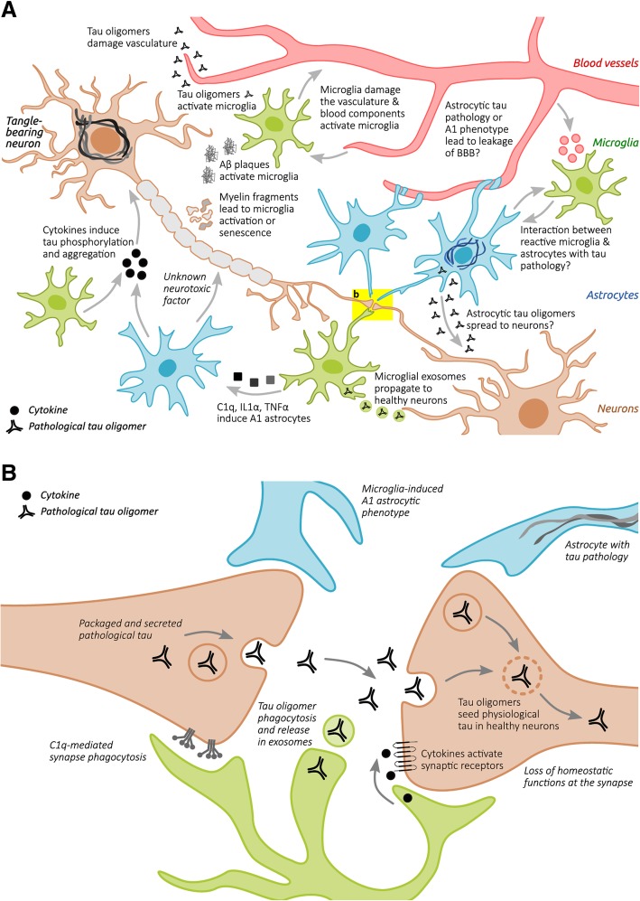
Tauopathies are a heterogenous class of diseases characterized by cellular accumulation of aggregated tau and include diseases such as Alzheimer's disease (AD), progressive supranuclear palsy and chronic traumatic encephalopathy. Tau pathology is strongly linked to neurodegeneration and clinical symptoms in tauopathy patients. Furthermore, synapse loss is an early pathological event in tauopathies and is the strongest correlate of cognitive decline. Tau pathology is additionally associated with chronic neuroinflammatory processes, such as reactive microglia, astrocytes, and increased levels of pro-inflammatory molecules (e.g. complement proteins, cytokines). Recent studies show that as the principal immune cells of the brain, microglia play a particularly important role in the initiation and progression of tau pathology and associated neurodegeneration. Furthermore, AD risk genes such as Triggering receptor expressed on myeloid cells 2 (TREM2) and Apolipoprotein E (APOE) are enriched in the innate immune system and modulate the neuroinflammatory response of microglia to tau pathology. Microglia can play an active role in synaptic dysfunction by abnormally phagocytosing synaptic compartments of neurons with tau pathology. Furthermore, microglia are involved in synaptic spreading of tau - a process which is thought to underlie the progressive nature of tau pathology propagation through the brain. Spreading of pathological tau is also the predominant target for tau-based immunotherapy. Active tau vaccines, therapeutic tau antibodies and other approaches targeting the immune system are actively explored as treatment options for AD and other tauopathies. This review describes the role of microglia in the pathobiology of tauopathies and the mechanism of action of potential therapeutics targeting the immune system in tauopathies.
Keywords: APOE4; Astrocytes; Complement; Microglia; Neurodegeneration; Neuroinflammation; Synaptic dysfunction; TREM2; Tau immunotherapy; Tau pathology.
Conflict of interest statement
TV and TH are employees of Axon Neuroscience R&D Services
Figures
Similar articles
-
Enhancing TREM2 expression activates microglia and modestly mitigates tau pathology and neurodegeneration.J Neuroinflammation. 2025 Mar 23;22(1):93. doi: 10.1186/s12974-025-03420-8. J Neuroinflammation. 2025. PMID: 40122810 Free PMC article.
-
Selective removal of astrocytic APOE4 strongly protects against tau-mediated neurodegeneration and decreases synaptic phagocytosis by microglia.Neuron. 2021 May 19;109(10):1657-1674.e7. doi: 10.1016/j.neuron.2021.03.024. Epub 2021 Apr 7. Neuron. 2021. PMID: 33831349 Free PMC article.
-
TREM2-Deficient Microglia Attenuate Tau Spreading In Vivo.Cells. 2023 Jun 10;12(12):1597. doi: 10.3390/cells12121597. Cells. 2023. PMID: 37371067 Free PMC article.
-
Glial contributions to neurodegeneration in tauopathies.Mol Neurodegener. 2017 Jun 29;12(1):50. doi: 10.1186/s13024-017-0192-x. Mol Neurodegener. 2017. PMID: 28662669 Free PMC article. Review.
-
Microglia in Alzheimer's Disease in the Context of Tau Pathology.Biomolecules. 2020 Oct 14;10(10):1439. doi: 10.3390/biom10101439. Biomolecules. 2020. PMID: 33066368 Free PMC article. Review.
Cited by
-
Neuroprotective Effect of SGLT2 Inhibitors.Molecules. 2021 Nov 28;26(23):7213. doi: 10.3390/molecules26237213. Molecules. 2021. PMID: 34885795 Free PMC article. Review.
-
Aggregated Tau-PHF6 (VQIVYK) Potentiates NLRP3 Inflammasome Expression and Autophagy in Human Microglial Cells.Cells. 2021 Jun 30;10(7):1652. doi: 10.3390/cells10071652. Cells. 2021. PMID: 34209408 Free PMC article.
-
Neuroinflammation and Tau Colocalize in vivo in Progressive Supranuclear Palsy.Ann Neurol. 2020 Dec;88(6):1194-1204. doi: 10.1002/ana.25911. Epub 2020 Oct 10. Ann Neurol. 2020. PMID: 32951237 Free PMC article.
-
Identification of retinal oligomeric, citrullinated, and other tau isoforms in early and advanced AD and relations to disease status.Acta Neuropathol. 2024 Jul 9;148(1):3. doi: 10.1007/s00401-024-02760-8. Acta Neuropathol. 2024. PMID: 38980423 Free PMC article.
-
The Role of Microglia in Perioperative Neurocognitive Disorders.Front Cell Neurosci. 2020 Aug 18;14:261. doi: 10.3389/fncel.2020.00261. eCollection 2020. Front Cell Neurosci. 2020. PMID: 32973455 Free PMC article. Review.
References
-
- Akiyoshi Ryohei, Wake Hiroaki, Kato Daisuke, Horiuchi Hiroshi, Ono Riho, Ikegami Ako, Haruwaka Koichiro, Omori Toshiaki, Tachibana Yoshihisa, Moorhouse Andrew J., Nabekura Junichi. Microglia Enhance Synapse Activity to Promote Local Network Synchronization. eneuro. 2018;5(5):ENEURO.0088-18.2018. doi: 10.1523/ENEURO.0088-18.2018. - DOI - PMC - PubMed
Publication types
MeSH terms
Substances
Grants and funding
LinkOut - more resources
Full Text Sources
Miscellaneous
