. 2019 Jul 19;8(7):bio039040.
doi: 10.1242/bio.039040.

Intestinal lipid droplets as novel mediators of host-pathogen interaction in Drosophila

Affiliations

Intestinal lipid droplets as novel mediators of host-pathogen interaction in Drosophila

Sneh Harsh et al. Biol Open. .

Abstract

Lipid droplets (LDs) are lipid-carrying multifunctional organelles, which might also interact with pathogens and influence the host immune response. However, the exact nature of these interactions remains currently unexplored. Here we show that systemic infection of Drosophila adult flies with non-pathogenic Escherichia coli, the extracellular bacterial pathogen Photorhabdus luminescens or the facultative intracellular pathogen Photorhabdus asymbiotica results in intestinal steatosis marked by lipid accumulation in the midgut. Accumulation of LDs in the midgut also correlates with increased whole-body lipid levels characterized by increased expression of genes regulating lipogenesis. The lipid-enriched midgut further displays reduced expression of the enteroendocrine-secreted hormone, Tachykinin. The observed lipid accumulation requires the Gram-negative cell wall pattern recognition molecule, PGRP-LC, but not PGRP-LE, for the humoral immune response. Altogether, our findings indicate that Drosophila LDs are inducible organelles, which can serve as markers for inflammation and, depending on the nature of the challenge, they can dictate the outcome of the infection.

Keywords: Drosophila; Infection; Lipid droplets; Midgut.


Conflict of interest statement

Competing interestsThe authors declare no competing or financial interests.

Figures

Fig. 1.
Fig. 1.
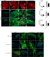
Systemic bacterial infection results in midgut lipid accumulation and increased fly body lipid storage. (A) Overview of the experimental workflow. Drosophila melanogaster background strain (strain w1118) flies were injected with PBS, E. coli, P. asymbiotica or P. luminescens, and fat body and midgut tissues were dissected to examine the status of LDs at 50, 30 and 24 hpi. (B) Representative images of fat body LDs from w1118 flies injected with PBS or 100–300 CFU of E. coli, P. asymbiotica or P. luminescens. Injection with PBS served as negative control. There was no substantial difference in the size of LDs among the different types of bacterial infections compared to PBS-injected controls. Fat body LDs were visualized with the fluorescent dye Nile Red (red) and nuclei were tagged with DAPI (blue). (C) Representative images of midgut LDs from w1118 flies injected with PBS or 100–300 CFU of E. coli, P. asymbiotica or P. luminescens. Bacterial infection resulted in dramatic accumulation of LDs in the midgut of the infected flies compared to PBS-injected controls. LDs were visualized with Nile Red (green) and nuclei with DAPI (blue). Lower panels show enlarged view of midgut LDs (outlined). (D–F) Systemic infection of background control flies with E. coli, P. asymbiotica or P. luminescens increased triglyceride levels in the fly body. Data represent the mean±s.d of three independent experiments. Asterisks indicate statistically significant differences compared to PBS-injected controls (Student's unpaired t-test, *P<0.05 and **P=0.005; ns, not significant). Scale bars: 100 μm.
Fig. 2.
Fig. 2.
Bacterial infection results in altered expression of genes regulating lipogenesis and lipolysis. Background control flies (strain w1118) were injected with 100–300 CFU of E. coli, P. asymbiotica or P. luminescens and then frozen at 50, 30 and 24 hpi, respectively. The infected flies were processed for transcript level analysis of lipid-metabolism related genes. PBS-injected flies served as negative control. (A–C) mRNA level of genes involved in lipogenesis. (D–F) Expression of lipolysis related genes in the whole fly. (A–C) Flies infected with E. coli, P. asymbiotica or P. luminescens showed consistent upregulation of genes involved in lipogenesis, marked by the increased expression of lipin and mdy. (D–F) Unlike lipogenesis, the effect on lipolysis was distinct among the different types of bacterial infection. lsd-1 and lsd-2 were used as read-outs for lipolysis. While lsd-1 was upregulated by E. coli, its level was reduced significantly upon infection with P. luminescens. lsd-2 was significantly and consistently upregulated upon infection with E. coli, P. asymbiotica or P. luminescens. All mRNA levels were normalized against RpL32 and three independent experiments were performed. Graphs depict the mean±s.d. Asterisks indicate statistically significant differences compared to PBS-injected controls (Student's unpaired t-test, *P<0.05, **P<0.005, ***P<0.001; ns, not significant).
Fig. 3.
Fig. 3.
Bacterial infection in Drosophila results in reduced expression of the gut-secreted hormone Tachykinin and insulin signaling. Background control flies (strain w1118) were injected with 100–300 CFU of E. coli, P. asymbiotica or P. luminescens and dissected gut tissues were examined for mRNA expression of gut-secreting hormones and insulin signaling. PBS-injected flies served as negative control. (A–C) mRNA levels of gut-secreting hormone TK. (D–F) Expression of 4E-BP and Impl2. (A–C) Flies infected with E. coli, P. asymbiotica or P. luminescens showed significantly decreased expression of TK as compared to the PBS-injected controls. TK-reduced levels of expression were consistent for all bacterial infections. (D–F) Gut tissues from flies infected with the pathogens P. asymbiotica or P. luminescens showed significant upregulation of 4E-BP and Impl2, the negative regulators of insulin signaling. Infection with E. coli caused no altered expression of 4E-BP and Impl2. Levels of mRNA were normalized against RpL32 and three independent experiments were performed. Graphs depict the mean±s.d. Asterisks indicate statistically significant differences compared to PBS injected controls (Student's unpaired t-test, *P<0.05, **P=0.001; ns, not significant).
Fig. 4.
Fig. 4.
Knockdown of the Gram-negative bacterial-recognition protein PGRP-LC ameliorates the bacterial infection-induced gut lipid accumulation. (A) Representative images of fat body LDs from background control flies (strain w1118) injected with PBS or 100–300 CFU of heat-inactivated E. coli, P. asymbiotica or P. luminescens. Injection with PBS served as negative control. There was no noticeable difference in the size of LDs between treatments. Fat body LDs were visualized with the fluorescent dye Nile Red (red) and nuclei were stained with DAPI (blue). (B) Midgut tissues from flies injected with PBS or heat-inactivated E. coli, P. asymbiotica or P. luminescens. Midgut tissues from flies injected with heat-inactivated bacteria showed marked accumulation of LDs as compared to PBS-injected controls. Midgut LDs were visualized with Nile Red (green) and nuclei with DAPI (blue). Lower panels show the enlarged view of midgut LDs (outlined). (C) qRT-PCR revealed increased expression of genes regulating lipogenesis, lipin and mdy in flies injected with heat-inactivated E. coli, P. asymbiotica or P. luminescens. (D) Representative images of midgut LDs from background control flies and flies mutant for PGRP-LE (yw PGRP-LE112), PGRP-LC (w; PGRP-LCΔE) upon injection with PBS, E. coli, P. asymbiotica or P. luminescens. Similar to the background controls (examined in both yw and w1118 strains, but for simplicity representative images from w1118 strain only are shown), PGRP-LE mutants showed dramatic accumulation of LDs in the midgut. In contrast, midgut tissues from PGRP-LC mutants did not show bacterial infection-induced lipid droplet accumulation following injection with E. coli, P. asymbiotica or P. luminescens. Midgut LDs were visualized with Nile Red (green) and nuclei were stained with DAPI (blue). Levels of mRNA were normalized against RpL32 and three independent experiments were performed. Graphs show the mean±s.d. Asterisks indicate statistically significant differences compared to PBS-injected controls (Student's unpaired t-test, ****P<0.0001, ***P<0.05, **P=0.0023, *P<0.05). Scale bars: 100 μm.
Fig. 5.
Fig. 5.
Intestinal steatosis modulates the survival of bacterially infected flies without affecting bacterial load. Survival and bacterial burden in flies knocked down for gut specific hormone TK driven under TKg-Gal4 (TKg>UAS-TK RNAi) following intrathoracic injection with 100–300 CFU of P. asymbiotica or P. luminescens. Injection with PBS served as negative control. (A,B) TK-silenced flies (TKg>UAS-TK RNAi) survived longer as compared to control flies (TKg>UAS-w RNAi) when challenged with P. asymbiotica. While control flies reached 50% survival by 30 hpi, TK-silenced flies reached 50% survival by 40 hpi. (B) Quantification of bacterial burden in control flies (TKg>UAS-w RNAi) and flies with knocked down TK (TKg>UAS-TK RNAi) upon infection with P. asymbiotica (40 hpi). (C,D) TK-silenced flies (TKg>UAS-TK RNAi) were more sensitive to P. luminescens and succumbed at a faster rate as compared to the controls (TKg>UAS-w RNAi). Survival of TK-silenced flies and control flies dropped to 50% at 18 hpi and 24 hpi with P. luminescens, respectively. (D) Quantification of bacterial burden in control flies and flies with knocked-down TK upon systemic infection with P. luminescens (18 hpi). Log-rank (Mantel-Cox) was used to analyze the data (***P<0.0001). CFU were determined by qRT-PCR of 16SrRNA against a standard bacterial curve and normalized against control flies (TKg>UAS-w RNAi). Three independent experiments were performed. Graphs show the mean±s.d. Statistical analysis was performed using Student's unpaired t-test (ns, not significant).
Fig. 6.
Fig. 6.
Adult immune pathway activation results in localized enlarged fat body LDs and non-autonomous midgut lipid accumulation. Toll and Imd signaling were constitutively activated in adult D. melanogaster and LD perturbation in the fat body and midgut were examined. Toll and Imd signaling were induced using the overexpression of activated Toll receptor UAS-Toll10b and overexpression of Relish (UAS-rel) under adult female fat body-specific driver Yolk-Gal4 (Yolk>UAS-Toll10b and Yolk>UAS-rel), respectively. (A) Representative images of adult fat body LDs for the indicated immune signaling. LDs were marked with the fluorescent dye Nile Red (red), and nuclei with DAPI (blue). Adult flies with upregulated Toll or Imd signaling showed strikingly enlarged LDs in the fat body as compared to the control Yolk-Gal4 strain. (B) Representative images of midgut LDs from flies carrying Yolk-Gal4, Yolk-Gal4-driven Toll or Imd overexpression (FB>UAS-Toll10b, FB>UAS-rel). Midgut tissues from adult flies overexpressing immune signaling pathways showed markedly increased accumulation of LDs compared to the control adult carrying Yolk-Gal4 alone. LDs were visualized with Nile Red (green) and nuclei with DAPI (blue). (C) Quantification of fat body LD size in flies overexpressing immune signaling pathways. (D) qRT-PCR analysis showing increased transcript levels of lipogenesis-regulating genes lipin and mdy in the adult flies carrying overexpression of immune signaling pathways. Levels of mRNA were normalized against RpL32 and three independent experiments were performed. Graphs depict the mean±s.d. Asterisks indicate statistically significant differences upon activation of immune signaling compared to Yolk-Gal4 (Student's unpaired t-test, *P<0.05, **P<0.005). Scale bars: 100 μm.
Fig. 7.
Fig. 7.
PGRP-LC-mediated intestinal steatosis confers a protective or harmful effect in flies responding to bacterial infection. (Upper panel) Scheme representing the host lipid dynamics in uninfected flies. LDs (red) are mainly localized in the fat body (Fb) and partly in proventriculus (pv) and midgut (mg) region of the gut. Gut hormone TK regulates lipid homeostasis in the gut as well as at the systemic level by suppressing lipogenesis. (Lower panel) Scheme representing the sequence of events triggered upon systemic bacterial infection. DAP-type peptidoglycan of Gram-negative bacteria is recognized by PGRP-LC, and this event is transduced in the form of intestinal steatosis marked by accumulation of LDs in the midgut of the infected flies without affecting fat body LDs. Intestinal steatosis is associated with reduced expression of TK, which in turn leads to increased rate of lipogenesis. The triggered intestinal steatosis can induce a protective or harmful response to the flies depending on the nature of bacterial infection.

References

    1. Anand P., Cermelli S., Li Z., Kassan A., Bosch M., Sigua R., Huang L., Ouellette A. J., Pol A., Welte M. A. et al. (2012). A novel role for lipid droplets in the organismal antibacterial response. Elife 1, e00003 10.7554/eLife.00003 - DOI - PMC - PubMed
    1. Araujo-Santos T., Rodriguez N. E., Moura-Pontes S., Dixt U. G., Abanades D. R., Bozza P. T., Wilson M. E. and Borges V. M. (2014). Role of prostaglandin F2alpha production in lipid bodies from Leishmania infantum chagasi: insights on virulence. J. Infect. Dis. 210, 1951-1961. 10.1093/infdis/jiu299 - DOI - PMC - PubMed
    1. Arrese E. L. and Soulages J. L. (2010). Insect fat body: energy, metabolism, and regulation. Annu. Rev. Entomol. 55, 207-225. 10.1146/annurev-ento-112408-085356 - DOI - PMC - PubMed
    1. Aymeric J. L., Givaudan A. and Duvic B. (2010). Imd pathway is involved in the interaction of Drosophila melanogaster with the entomopathogenic bacteria, Xenorhabdus nematophila and Photorhabdus luminescens. Mol. Immunol. 47, 2342-2348. 10.1016/j.molimm.2010.05.012 - DOI - PubMed
    1. Barletta A. B., Alves L. R., Silva M. C., Sim S., Dimopoulos G., Liechocki S., Maya-Monteiro C. M. and Sorgine M. H. (2016). Emerging role of lipid droplets in Aedes aegypti immune response against bacteria and Dengue virus. Sci. Rep. 6, 19928 10.1038/srep19928 - DOI - PMC - PubMed

LinkOut - more resources