. 2019 Jul 5;9(1):9721.
doi: 10.1038/s41598-019-46144-5.

A novel motif in the proximal C-terminus of Pannexin 1 regulates cell surface localization

Affiliations

A novel motif in the proximal C-terminus of Pannexin 1 regulates cell surface localization

Anna L Epp et al. Sci Rep. .

Abstract

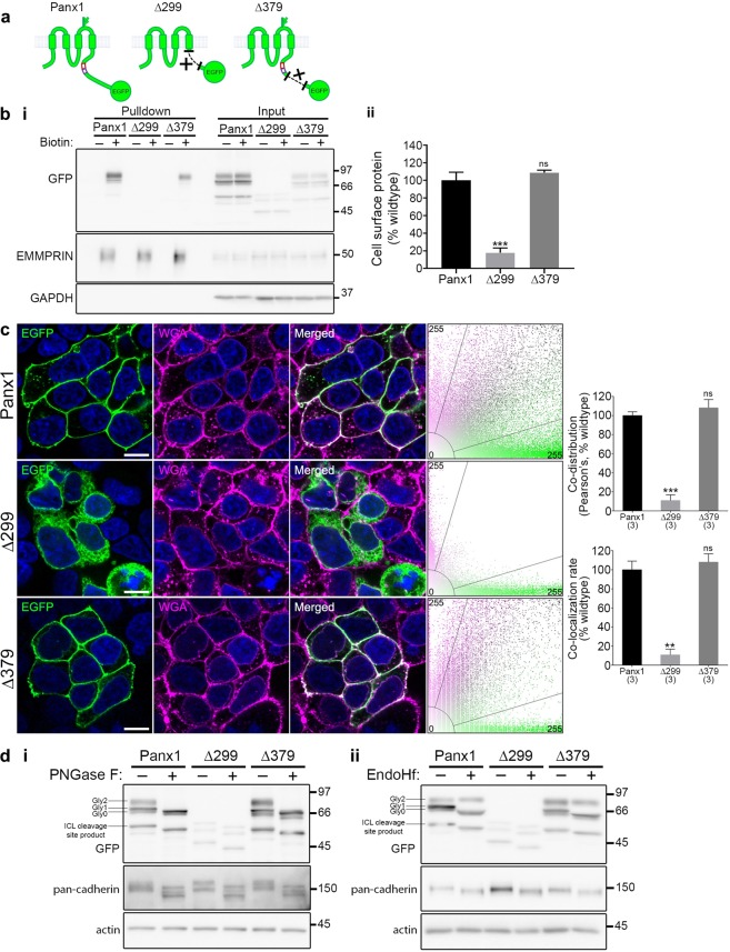
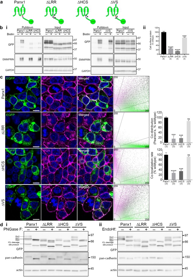
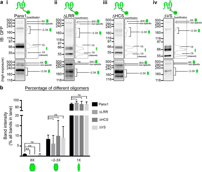
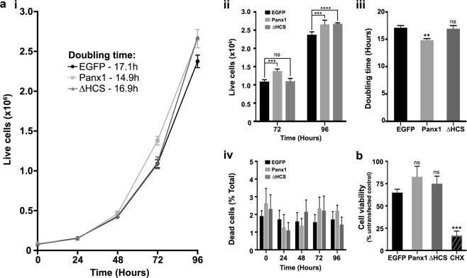
The Pannexin 1 (Panx1) ion and metabolite channel is expressed in a wide variety of cells where it regulates a number of cell behaviours including proliferation and differentiation. Panx1 is expressed on the cell surface as well as intracellular membranes. Previous work suggests that a region within the proximal Panx1 C-terminus (Panx1CT) regulates cell surface localization. Here we report the discovery of a putative leucine-rich repeat (LRR) motif in the proximal Panx1CT necessary for Panx1 cell surface expression in HEK293T cells. Deletion of the putative LRR motif results in significant loss of Panx1 cell surface distribution. Outcomes of complementary cell surface oligomerization and glycosylation state analyses were consistent with reduced cell surface expression of Panx1 LRR deletion mutants. Of note, the oligomerization analysis revealed the presence of putative dimers and trimers of Panx1 at the cell surface. Expression of Panx1 increased HEK293T cell growth and reduced doubling time, while expression of a Panx1 LRR deletion mutant (highly conserved segment) did not reproduce this effect. In summary, here we discovered the presence of a putative LRR motif in the Panx1CT that impacts on Panx1 cell surface localization. Overall these findings provide new insights into the molecular mechanisms underlying C-terminal regulation of Panx1 trafficking and raise potential new lines of investigation with respect to Panx1 oligomerization and glycosylation.


Conflict of interest statement

The authors declare no competing interests. A disclosure has been made with the University of Victoria Research Partnerships and Knowledge Mobilization, and a provisional patent application has been filed for a peptide targeting a Panx1-Crmp2 interaction (US Application No. 62/767,806) that does not relate directly to this study.

Figures

Figure 1
Figure 1
The proximal Panx1CT is necessary for cell surface localization of Panx1-EGFP. (a) Schematic of full length Panx1-EGFP and the Panx1∆299-EGFP and Panx1∆379-EGFP deletion mutants. (b) Cell surface biotinylation assays reveal that the proximal Panx1CT is required for cell surface localization in HEK293T cells. (i) Representative Western blot of pulldown (cell surface protein) and input. Anti-EMMPRIN was used as a positive control for biotin pulldown and as a loading control, and anti-GAPDH was used as a negative control against biotin internalization. (ii) Panx1∆299-EGFP exhibited reduced cell surface levels compared to Panx1-EGFP, while surface levels of Panx1∆379-EGFP were similar to those of Panx1-EGFP. Data are presented as mean ± SEM. One-way ANOVA with Dunnett’s multiple comparisons test, N = 3, α = 0.05, F(2,6) = 62.39, **P = 0.0002, ns, non-significant. (c) Representative confocal micrographs of HEK293T cells overexpressing Panx1-EGFP, Panx1∆379-EGFP, or Panx1∆299-EGFP (green). Hoechst was used as a nuclear counterstain (blue) and wheat-germ agglutinin (WGA) was used as a plasma membrane marker (magenta). Overlapping EGFP and WGA signals (white) and scatterplots with EGFP and WGA fluorescence signals show the co-distriution of these proteins along the cell membrane. Panx1-EGFP and Panx1∆379-EGFP co-distributed with WGA, while Panx1∆299-EGFP did not. One-way ANOVA with Dunnett’s multiple comparison test, N = 3, α = 0.05; Pearson’s: F(2,6) = 62.29, ***P = 0.0002, ns, non-significant; Colocalization rate: F(2,6) = 26.81, **P = 0.0014, ns, non-significant. Scale bars, 10 μm. Data are presented as mean ± SEM. (d) Deglycosylation assays using (i) PNGase F or (ii) EndoHf reveal Panx1-EGFP and Panx1∆379-EGFP exhibited Gly0, Gly1, and Gly2 glycosylation species, while Panx1∆299-EGFP exhibited only Gly0 and Gly1 forms. Anti-pan-cadherin and anti-β-actin were used as a positive and negative controls for deglycosylation, respectively. This figure was modified from Epp 2019. For uncropped images of all Western blots in this figure, please see Supplementary Fig. S3.
Figure 2
Figure 2
Surface oligomerization is preserved with deletion of the distal Panx1CT. (a) Crosslinking assays reveal oligomerization profiles of C-terminal deletion mutants. Representative Western blots observing HEK293T cell lysates post-transfection with (i) Panx1-EGFP, (ii) Panx1∆379-EGFP, or (iii) Panx1∆299-EGFP, and after crosslinking with the cell-impermeable crosslinker, BS3, to observe surface-localized oligomers (6× or ~2–3×). The plot of each quantification is included to shed light on the analytical process. An antibody for Crmp2, an intracellular protein known to form tetramers, was used as a negative control to ensure BS3 had not entered the cell. Crmp2 oligomerization did not increase in the presence of BS3, as expected. (b) Each oligomeric band was quantified and expressed as a percentage of the entire GFP signal in each lane. Non-specific bands were excluded from the analysis. Mutants lacking the distal Panx1CT (Panx1∆379-EGFP) had the same oligomerization profiles as full length Panx1-EGFP. Data are presented as mean ± SEM. One-way ANOVA with Dunnett’s multiple comparisons test, N = 3, α = 0.05; F(2,6) = 17.59, **P = 0.0023 (6×); F(2,6) = 15.73, *P = 0.0127 (3×); F(2,6) = 13.84, *P = 0.0169 (1×); ns, non-significant. All samples were derived from the same experiment and processed in parallel. This figure was modified from Epp 2019. For uncropped images of all Western blots in this figure, please see Supplementary Fig. S4
Figure 3
Figure 3
The Panx1CT amino acid sequence contains 5 LRR HCS consensus sequences. Sequence alignment of Panx1CT from human, mouse, and rat. Each of the identified HCS regions are indicated in red, and labeled with the corresponding consensus sequence. The full LRR identified by ScanProsite was separated into an HCS and VS (the VS is indicated in purple). The alignment was generated using CLUSTAL O (1.2.3) with NCBI accession numbers: NP_056183.2 (human), NP_062355.2 (mouse), and NP_955239.1 (rat). This figure was modified from Epp 2019.
Figure 4
Figure 4
A novel putative LRR motif in the Panx1CT is necessary for trafficking Panx1-EGFP to the cell surface. (a) Schematic of full length Panx1-EGFP and the Panx1∆LRR-EGFP, Panx1∆HCS-EGFP, and Panx1∆VS-EGFP deletion mutants. (b) Cell surface biotinylation assays reveal that the putative LRR motif, or HCS alone, is required for cell surface localization, while the VS is also mildly important. (i) Representative Western blot of pulldown (cell surface protein) and input. Anti-EMMPRIN was used as a positive control for biotin pulldown and as a loading control, and anti-GAPDH was used as a negative control against biotin internalization. (ii) Panx1∆LRR-EGFP and Panx1∆HCS-EGFP exhibited markedly reduced cell surface levels compared to Panx1-EGFP, while Panx1∆VS-EGFP exhibited a relatively small reduction cell surface levels compared to Panx1-EGFP. Data are presented as mean ± SEM. One-way ANOVA with Dunnett’s multiple comparisons test, N = 3, α = 0.05, F(3, 11) = 152.4, ***P = 0.0007, ****P < 0.0001, ns, non-significant. (c) Confocal images of HEK293T cells overexpressing Panx1-EGFP, Panx1∆LRR-EGFP, Panx1∆HCS-EGFP, or Panx1∆VS-EGFP (green). Hoechst was used as a nuclear counterstain (blue) and wheat-germ agglutinin (WGA) was used as a plasma membrane marker (magenta). Overlapping EGFP and WGA signals (white) and scatterplots with EGFP and WGA fluorescence signals show the co-distribution of these proteins along the cell membrane. While both Panx1-EGFP and Panx1∆VS-EGFP co-distributed with WGA, Panx1∆LRR-EGFP and Panx1∆HCS-EGFP did not. N = 3, α = 0.05; Pearson’s: F(2,6) = 32.94, ***P = 0.0002, ns, non-significant; Colocalization rate: F(2,6) = 38.99, ***P = 0.0002, ****P < 0.0001, ns, non-significant. Scale bars, 10 μm. Data are presented as mean ± SEM. (d) Deglycosylation assays using (i) PNGase F or (ii) EndoHf reveal Panx1-EGFP and Panx1∆VS-EGFP exhibited Gly0, Gly1, and Gly2 glycosylation species, while Panx1∆LRR-EGFP and Panx1∆HCS-EGFP exhibited only Gly0 and Gly1 forms. Anti-pan-cadherin and anti-β-actin were used as a positive and negative controls for deglycosylation, respectively. This figure was modified from Epp 2019. For uncropped images of all Western blots in this figure, please see Supplementary Fig. S5.
Figure 5
Figure 5
Surface hexamers are not detected with complete putative LRR motif or HCS sequence deletion. (a) Crosslinking assays reveal oligomerization profiles of LRR motif deletion mutants. Representative Western blots observing HEK293T cell lysates post-transfection with (i) Panx1-EGFP, (ii) Panx1∆LRR-EGFP, (iii) Panx1∆HCS-EGFP, or (iv) Panx1∆VS-EGFP, and after crosslinking with the cell-impermeable crosslinker, BS3, to observe surface-localized oligomers (6× or ~2–3×). Higher exposures of the regions (above 116 kDa) were included to better illustrate the presence or absence of high molecular weight bands. (b) Individual oligomeric bands were quantified and expressed as a percentage of the entire GFP signal in each lane. Non-specific bands were excluded from the analysis. Data are presented as mean ± SEM. One-way ANOVA with Dunnett’s multiple comparisons test, N = 3, α = 0.05, F(3,8) = 10.59, **P = 0.0072 (6×), ns, non-significant. All samples were derived from the same experiment and processed in parallel. This figure was modified from Epp 2019. For uncropped images of all Western blots in this figure, please see Supplementary Fig. S6.
Figure 6
Figure 6
Panx1-EGFP overexpression increases HEK293T cell proliferation, but not when it lacks the HCS sequence (Panx1∆HCS-EGFP). (a) Trypan blue proliferation assays. (i) HEK293T cells overexpressing either Panx1-EGFP, Panx1∆HCS-EGFP, or EGFP control were counted at 5 time points with the first time point, T0, taking place at 24 h post-transfection (6 h after re-plating). Cell counts were used to plot growth curves. (ii) Histogram with cell counts from T0, T72, and T96. Data are presented as mean ± SEM. Two-way ANOVA with Dunnett’s multiple comparisons test, N = 4, α = 0.05, F(2,45) = 8.459, ***P = 0.0002, ****P < 0.0001, ns, non-significant. (iii) Doubling time comparisons of each condition, calculated using data from time points in the exponential growth phase. (iv) Dead cell counts at each time point, expressed as a percent of total cells. There were no significant differences in dead cell populations. Data are presented as mean ± SEM. One-way ANOVA with Dunnett’s multiple comparisons test, N = 4, α = 0.05, F(2,9) = 8.681, **P = 0.0079, ns, non-significant. (b) MTT assays performed at T72 on HEK293T cells overexpressing either EGFP, Panx1-EGFP, Panx1∆HCS-EGFP, or untransfected cells treated with a toxic dose (200 μg/mL) of cycloheximide (CHX) confirm that differences in cell numbers were not due to alterations in cell viability. Data are presented as the mean percentage of untransfected control ± SEM. One-way ANOVA with Dunnett’s multiple comparisons test, N = 6, α = 0.05, F(3,20) = 17.35, ****P < 0.0001, ns, non-significant. This figure was modified from Epp 2019.

References

    1. Boyce AKJ, Epp AL, Nagarajan A, Swayne LA. Transcriptional and post-translational regulation of pannexins. Biochimica et Biophysica Acta - Biomembranes. 2018;1860:72–82. doi: 10.1016/j.bbamem.2017.03.004. - DOI - PubMed
    1. Penuela S, et al. Pannexin 1 and pannexin 3 are glycoproteins that exhibit many distinct characteristics from the connexin family of gap junction proteins. J Cell Sci. 2007;120:3772–3783. doi: 10.1242/jcs.009514. - DOI - PubMed
    1. Boassa D, et al. Pannexin1 channels contain a glycosylation site that targets the hexamer to the plasma membrane. J Biol Chem. 2007;282:31733–31743. doi: 10.1074/jbc.M702422200. - DOI - PubMed
    1. Penuela S, Bhalla R, Nag K, Laird DW. Glycosylation regulates pannexin intermixing and cellular localization. Mol Biol Cell. 2009;20:4313–4323. doi: 10.1091/mbc.e09-01-0067. - DOI - PMC - PubMed
    1. Bhalla-Gehi R, Penuela S, Churko JM, Shao Q, Laird DW. Pannexin1 and pannexin3 delivery, cell surface dynamics, and cytoskeletal interactions. J Biol Chem. 2010;285:9147–9160. doi: 10.1074/jbc.M109.082008. - DOI - PMC - PubMed

Publication types