Review
. 2020 Feb;37(1):27-40.
doi: 10.1007/s10719-019-09874-2. Epub 2019 Jul 5.

Anionic and zwitterionic moieties as widespread glycan modifications in non-vertebrates

Affiliations
Review

Anionic and zwitterionic moieties as widespread glycan modifications in non-vertebrates

Katharina Paschinger et al. Glycoconj J. 2020 Feb.

Abstract

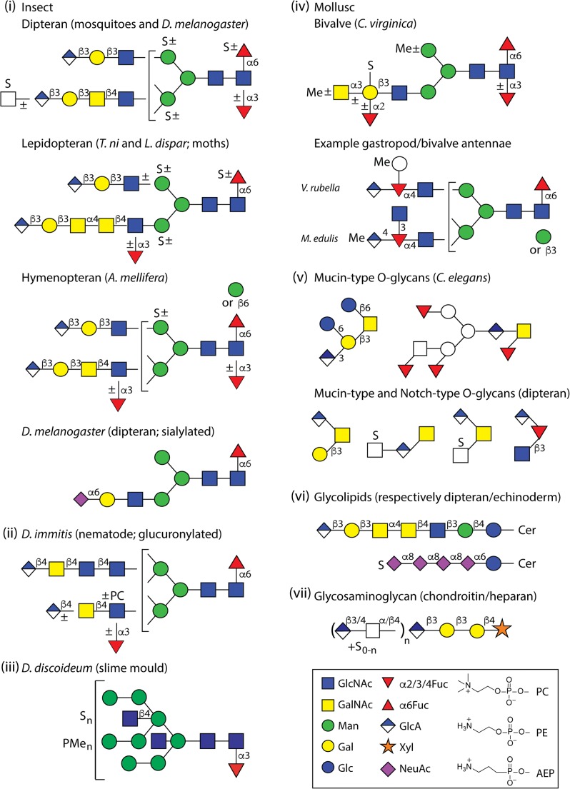
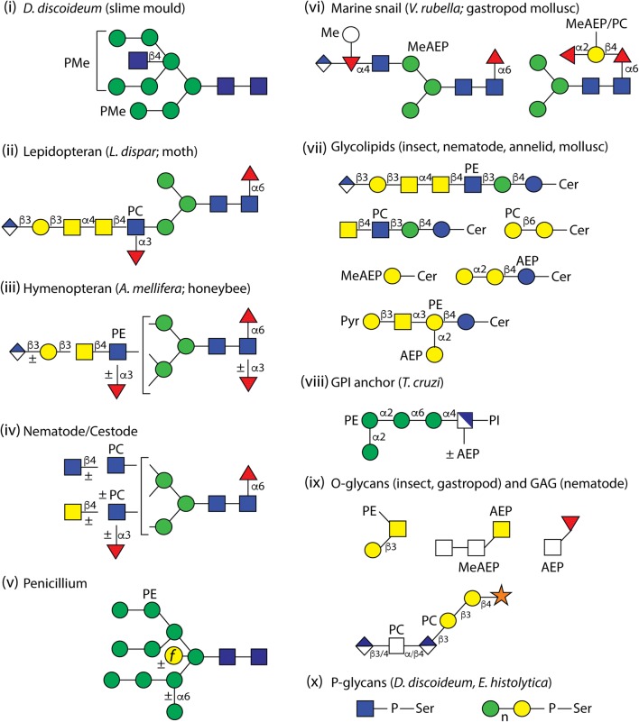
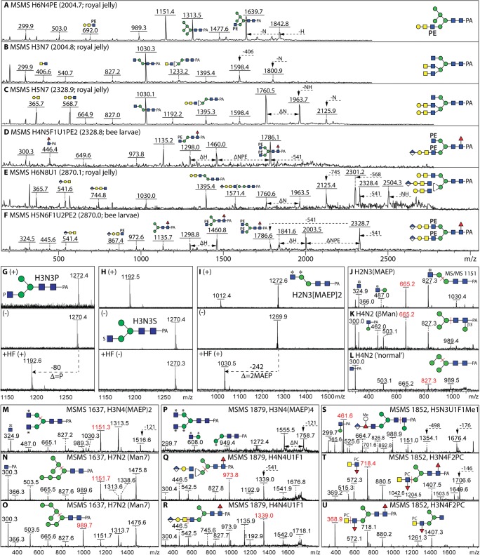
Glycan structures in non-vertebrates are highly variable; it can be assumed that this is a product of evolution and speciation, not that it is just a random event. However, in animals and protists, there is a relatively limited repertoire of around ten monosaccharide building blocks, most of which are neutral in terms of charge. While two monosaccharide types in eukaryotes (hexuronic and sialic acids) are anionic, there are a number of organic or inorganic modifications of glycans such as sulphate, pyruvate, phosphate, phosphorylcholine, phosphoethanolamine and aminoethylphosphonate that also confer a 'charged' nature (either anionic or zwitterionic) to glycoconjugate structures. These alter the physicochemical properties of the glycans to which they are attached, change their ionisation when analysing them by mass spectrometry and result in different interactions with protein receptors. Here, we focus on N-glycans carrying anionic and zwitterionic modifications in protists and invertebrates, but make some reference to O-glycans, glycolipids and glycosaminoglycans which also contain such moieties. The conclusion is that 'charged' glycoconjugates are a widespread, but easily overlooked, feature of 'lower' organisms.

Keywords: Glucuronic acid; Glycans; Glycomics; Insect; Mollusc; Nematode; Phosphoethanolamine; Phosphorylcholine; Sulphate.


Conflict of interest statement

The authors declare that they have no conflicts of interest.

Figures

Fig. 1
Fig. 1
Example glucuronylated, sialylated and sulphated glycans. Various features of N-glycans with anionic moieties are shown to highlight the variations from (i) dipteran (Aedes aegyptii, Anopheles gambiae and Drosophila melanogaster), lepidopteran (Trichoplusia ni and Lymantria dispar) and hymenopteran (Apis mellifera) insect species, (ii) a nematode (specifically Dirofilaria), (iii) the cellular slime mould Dictyostelium discoideum and (iv) Crassostrea virginica (oyster), Volvarina rubella (marine snail; upper depicted arm) and Mytilus edulis (blue mussel; lower depicted arm). Also shown are (v) example ‘mucin-type’ and ‘Notch-type’ O-glycans from nematodes and insects, (vi) glycolipids from Drosophila melanogaster (dipteran; glucuronylated) and Hemicentrotus pulcherrimus (echinoderm; sialylated) and (vii) the glycosaminoglycans (the latter being common to all animals). The glycans are depicted according to the Symbolic Nomenclature for Glycans (see box); MeAEP, methylaminoethylphosphonate; P, phosphate; PC, phosphorylcholine; PE, phosphoethanolamine; PMe, methylphosphate; Pyr, pyruvate, S, sulphate; white circles or boxes indicate undefined hexoses or N-acetylhexosamines. Linkages are defined for proven antennal motifs, while basic trimannosylchitobiosyl cores are assumed for all N-glycans
Fig. 2
Fig. 2
Example zwitterionic and phosphodiester structures. The depicted structures are (i) a methylphosphorylated N-glycan structure from Dictyostelium discoideum, (ii) an N-glycan from Lymantria dispar modified with glucuronic acid and phosphorylcholine, (iii) a schematic honeybee N-glycan, (iv) a schematic glycan from either nematode or cestode species, (v) a phosphoethanolamine-modified N-glycan from Penicillium spp. optionally with an ‘outer chain’ mannose and a bisecting galactofuranose, (vi) two N-glycans with methylaminoethylphosphonate or phosphorylcholine from Volvarina rubella, (vii) glycolipids from insects (example from Drosophila), nematodes and annelids (with phosphorylcholine) and molluscs (with aminoethylphosphonate; also observed with pyruvate and phosphoethanolamine modifications), (viii) a glycosylphosphatidylinositol (GPI) anchor from the Trypanosoma cruzi NETNES protein, (ix) a mucin-type O-glycan from wasp, a mucin-type and a Notch-type from Volvarina rubella, a glycosaminoglycan-like glycan from Oesophagostomum dentatum and (x) phospho-linked sugars from either Dictyostelium discoideum or Entamoeba histolytica. AEP, aminoethylphosphonate; MeAEP, methylaminoethylphosphonate; P, phosphate; PC, phosphorylcholine; PE, phosphoethanolamine; PMe, methylphosphate; Pyr, pyruvate
Fig. 3
Fig. 3
Mass spectrometry of isobaric/isomeric structures. a-f Example positive mode MS/MS of N-glycans from Apis mellifera royal jelly or larvae which isobarically differ depending on the presence of phosphoethanolamine, LacdiNAc or glucuronylated motifs. g-i Example positive (+) or negative (−) mode MS of isobaric glycans from an echinoderm or Volvarina with masses of 1271 Da carrying either phosphate, sulphate (note in-source loss in positive mode) or methylaminoethylphosphonate moieties and are distinguishable due to their ionisation in positive mode or sensitivity to hydrofluoric acid (HF). j-l Positive mode MS/MS of isobaric/isomeric variations of 1150 Da (m/z 1151) from Volvarina rubella either methylaminoethylphosphonate-modified, core β-mannosylated or ‘normal’ paucimannosidic. m-u Positive mode MS/MS of variations of glycans of m/z 1637, 1879 or 1852 which are either methylaminoethylphosphonate-modified (Volvarina), glucuronylated (Apis), phosphorylcholine-modified (Trichoplusia or Dirofilaria) or standard oligomannosidic structures. Annotated are the key fragments (symbolic nomenclature), losses (with arrows) or cleavages (red bars and m/z values); all glycans were reductively aminated with 2-aminopyridine (PA) which yields typical reducing terminal Y fragments of m/z 300, 446 or 462 (GlcNAc1Fuc0–1Man0–1-PA). Abbreviated compositions of the form HxNyF0–1 U0–1 correspond to HexxHexNAcyFuc0–1HexA0–1; MEAP (or *), methylaminoethylphosphonate; PC, phosphorylcholine; PE, phosphoethanolamine; S, sulphate

Similar articles

Cited by

References

    1. Varki A. Nothing in glycobiology makes sense, except in the light of evolution. Cell. 2006;126:841–845. doi: 10.1016/j.cell.2006.08.022. - DOI - PubMed
    1. Schauer R, Kamerling JP. Exploration of the sialic acid world. Adv. Carbohydr. Chem. Biochem. 2018;75:1–213. doi: 10.1016/bs.accb.2018.09.001. - DOI - PMC - PubMed
    1. Bülow HE, Hobert O. The molecular diversity of glycosaminoglycans shapes animal development. Annu. Rev. Cell Dev. Biol. 2006;22:375–407. doi: 10.1146/annurev.cellbio.22.010605.093433. - DOI - PubMed
    1. Morita I, Kizuka Y, Kakuda S, Oka S. Expression and function of the HNK-1 carbohydrate. J. Biochem. 2008;143:719–724. doi: 10.1093/jb/mvm221. - DOI - PubMed
    1. Honke K, Taniguchi N. Sulfotransferases and sulfated oligosaccharides. Med. Res. Rev. 2002;22:637–654. doi: 10.1002/med.10020. - DOI - PubMed

Publication types

LinkOut - more resources