. 2019 Oct:21:102049.
doi: 10.1016/j.nano.2019.102049. Epub 2019 Jul 3.

Evaluation of an adjuvanted hydrogel-based pDNA nanoparticulate vaccine for rabies prevention and immunocontraception

Affiliations

Evaluation of an adjuvanted hydrogel-based pDNA nanoparticulate vaccine for rabies prevention and immunocontraception

Amit Bansal et al. Nanomedicine. 2019 Oct.

Abstract

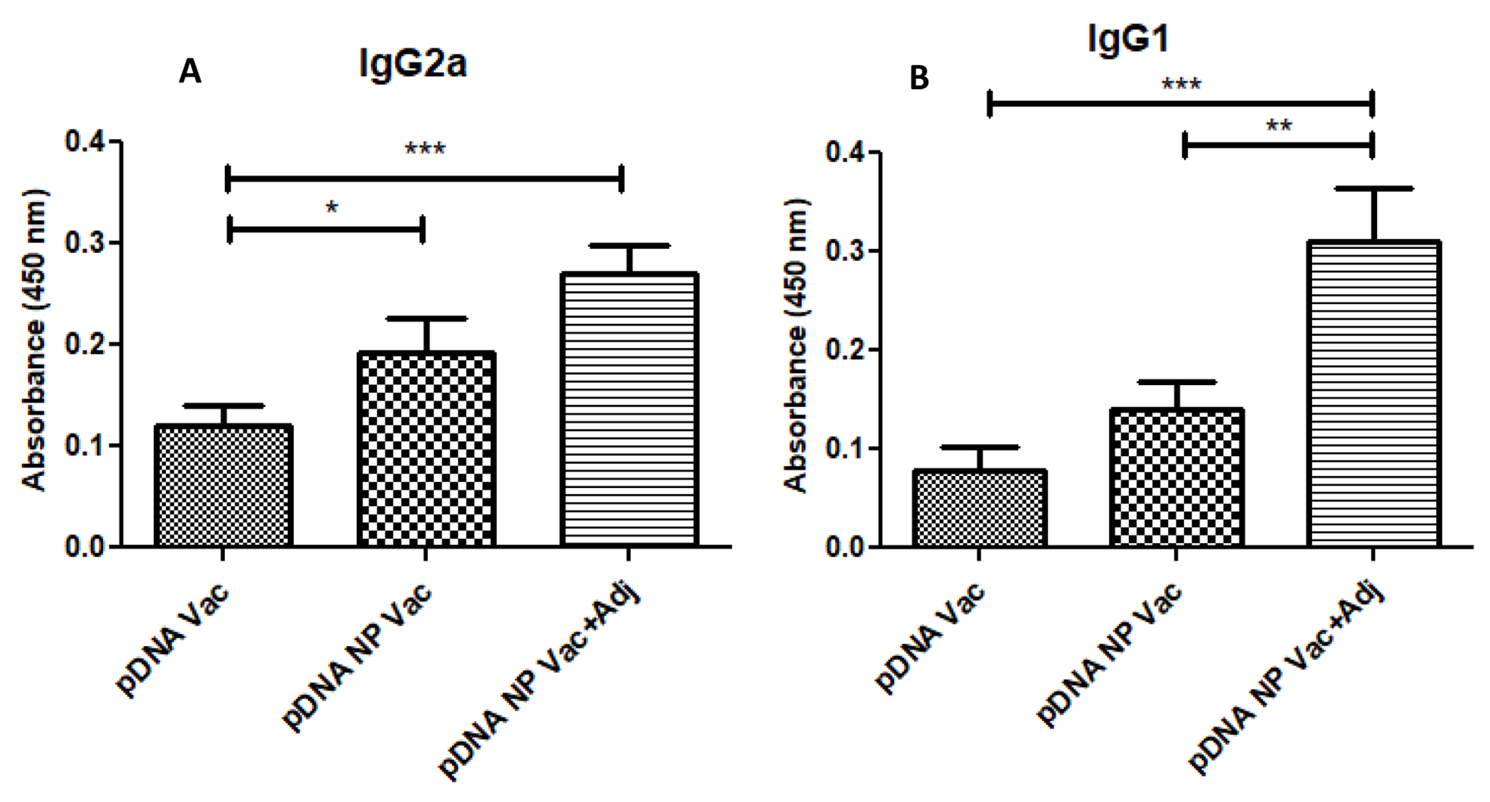
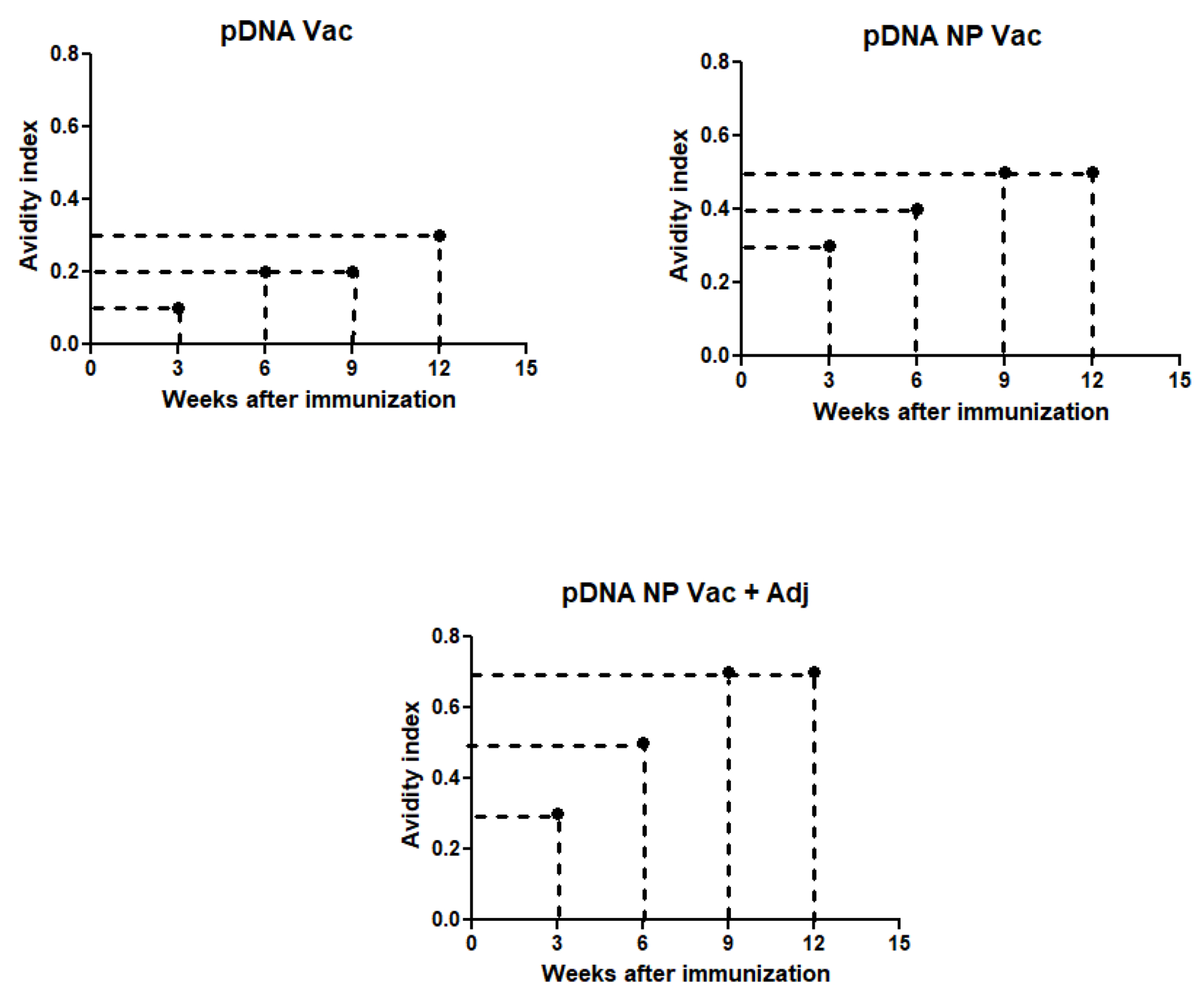
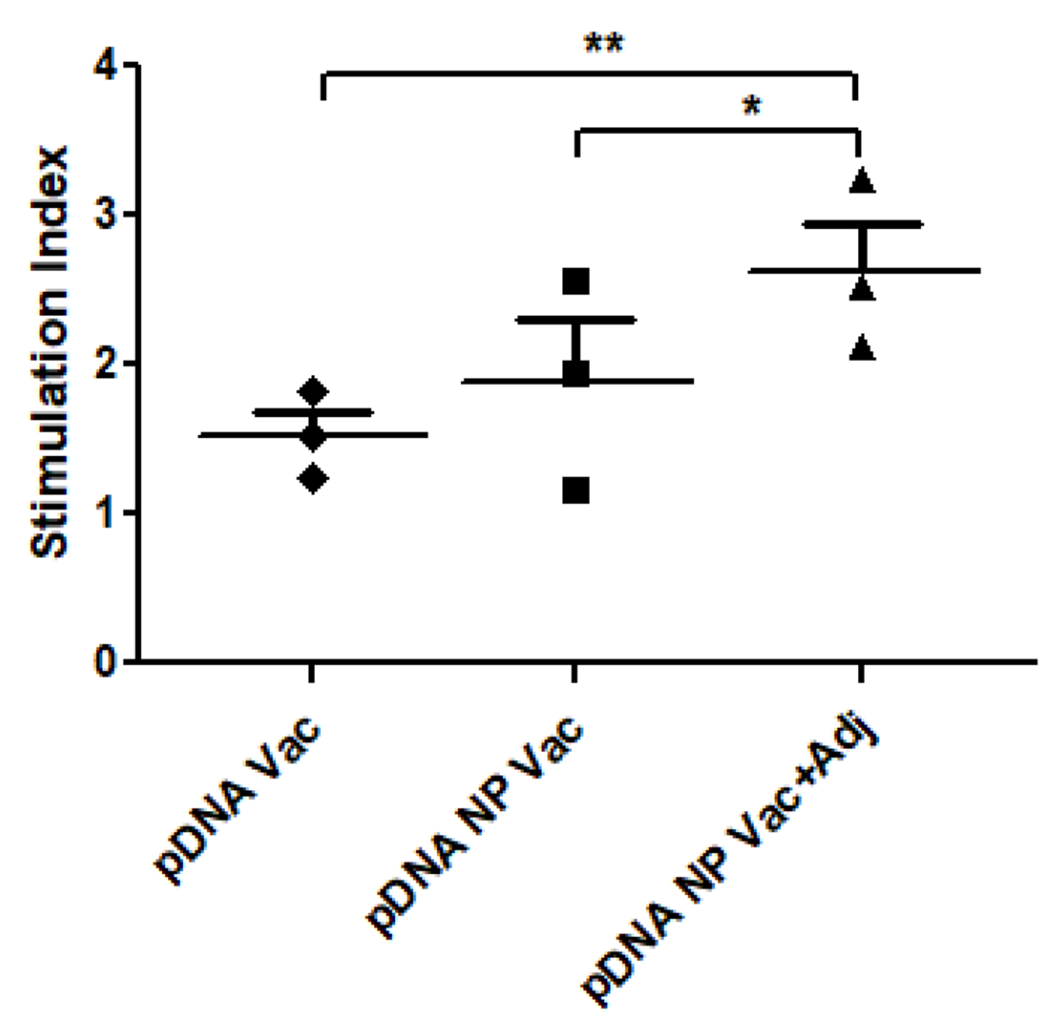
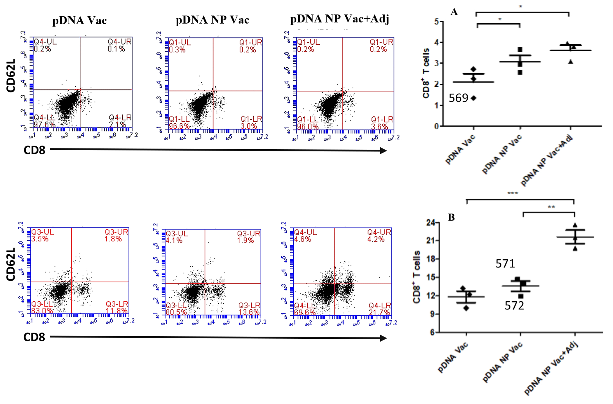
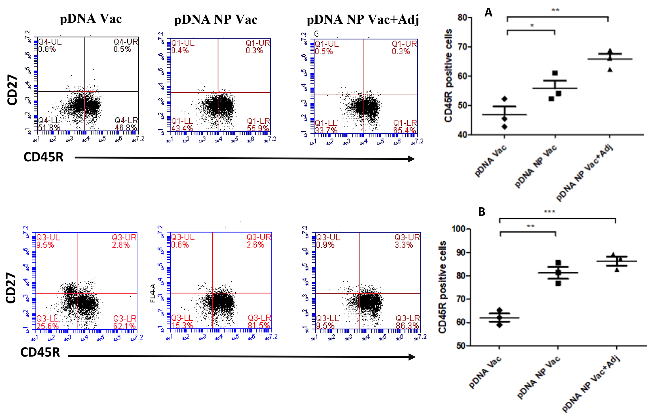
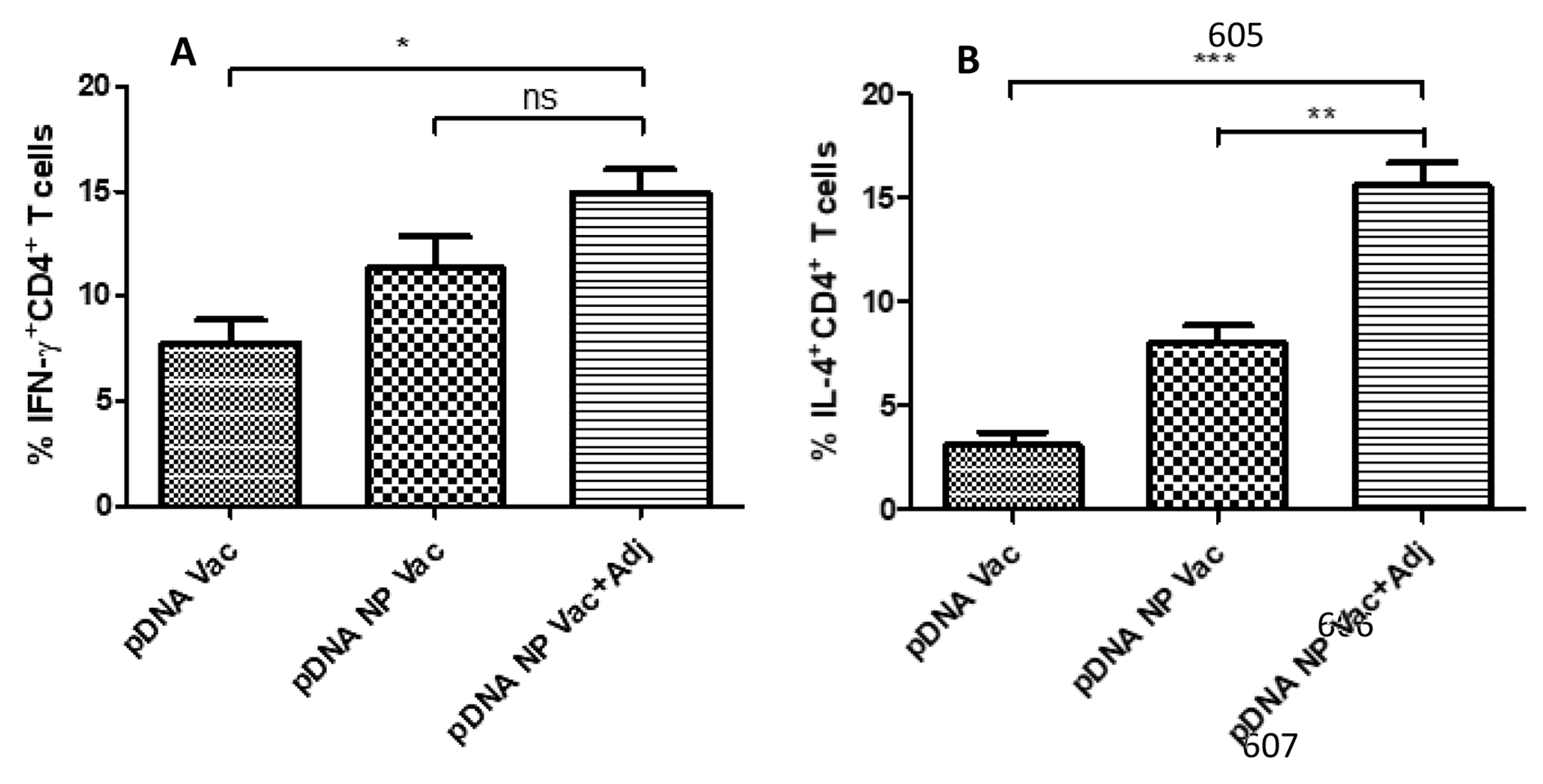
Immunocontraceptive vaccination is becoming an acceptable strategy in managing animal populations. Mass vaccination of dogs is the most cost-effective and efficient method to control rabies, and combination of rabies vaccination and animal population control will be an added advantage. In this study, we developed an adjuvanted hydrogel-based pDNA nanoparticulate vaccine for rabies protection and immunocontraception. In vivo, we observed an immune response skewed toward a Th2 type, in contrast to the Th1 type in our previous pDNA study. The observation was verified by the IgG2a/IgG1 ratio (<1), and cytokine expression profile of IL-4 and IFN-γ. The humoral immune response is key for rabies protection and a GnRH antibody-based immunocontraception. In mice, anti-GnRH antibody titers were detected 4 weeks after immunization and lasted for 12 weeks, post animal experiment was terminated. The adjuvanted pDNA nanoparticulate vaccine shows promise for future studies evaluating protection from rabies challenge and prevention of animal breeding.

Keywords: Gonadotrophin-releasing hormone (GnRH); Immunocontraceptive vaccine; Poloxamer gels; Rabies.


Conflict of interest statement

Disclosure

The authors report no conflicts of interest in this work. The authors alone are responsible for the content and writing of this article.

Figures

Figure 1 -
Figure 1 -
Ethidium bromide exclusion assay measuring the percent change in fluorescence intensity. (A) Percent decrease in fluorescence intensity was observed on addition of increasing concentrations of chitosan glutamate (0.1–0.5 μg/μL) and (B) percent decrease in fluorescence intensity with change in pDNA to nanoparticles ratio (P/N). ns - Not significant, *p<0.05 significant **p<0.01 very significant, ***p<0.001 extremely significant. Data are expressed as mean ± SEM.
Figure 2 -
Figure 2 -
Nitrite release from DC 2.4 cells was measured by Griess assay. Murine DC 2.4 cells were pulsed with LPS (5 μg) as positive control, pDNA (1 μg), blank NP (50 μg), pDNA nanoparticulate vaccine (50 μg, pDNA amount 1 μg), and pDNA nanoparticulate vaccine plus adjuvants Alum and MF59. There was a significant increase in nitrite release by groups received pDNA nanoparticulate vaccine with or without adjuvant Alum and MF59 (*p<0.05 significant, **p<0.01very significant, ***p<0.001 extremely significant) measured using one-way ANOVA with Tukey post hoc test. Data are expressed as mean ± SEM.
Figure 3 –
Figure 3 –
Upregulation of maturation markers (i.e., MHC I and MHC II) on DCs post-exposure to various treatments. The expression of co-stimulatory molecules was detected by flow cytometry analysis of fluorescence-labelled CD80 and CD40 antibodies. DC 2.4 cells were exposed to an equivalent amount of pDNA in all treatment groups. Mean fluorescence intensity (MFI) ratios of treatment samples were plotted to untreated samples. Results were analyzed using one-way ANOVA followed by post hoc Tukey’s multiple comparison test (*p<0.05 significant, **p<0.01 very significant, ***p<0.001 extremely significant). Data are expressed as mean±SEM, n=3, triplicate.
Figure 4 -
Figure 4 -
Antibody titers (IgG) in mice after rabies vaccination administered via intramuscular route (IM). Mice (n=6) were vaccinated on day 1 with pDNA (100 μg) in hydrogel, pDNA nanoparticulate vaccine in hydrogel, and pDNA nanoparticulate vaccine plus adjuvant in hydrogel. Blood was collected every week post-vaccination and analyzed for anti-GnRH antibodies using ELISA. Results were compared statistically among groups every week (*p<0.05 significant, **p<0.01very significant, ***p<0.001 extremely significant) using one-way ANOVA with Tukey post hoc test. Data are expressed as mean±SEM.
Figure 5 -
Figure 5 -
The anti-GnRH specific IgG subclass immune response. IgG subclass (IgG1, IgG2a) titers were measured in sera of mice (n=6) post vaccination on week 10. The IgG1/IgG2a ratio (ratios>1 and <1 indicate a Th2 and Th1 polarized response, respectively). A significant increase in titers was observed in pDNA nanoparticulate vaccine adjuvanted group (Alum and MF59). The p value was calculated (*p<0.05 significant, **p<0.01very significant, ***p<0.001 extremely significant) using one-way ANOVA with Tukey post hoc test. Data are expressed as mean ± SEM.
Figure 6 –
Figure 6 –
Avidity index was determined by incubating serum with increasing concentrations of sodium thiocyanate and calculated as 50% reduction in absorbance from untreated samples. The three groups used for the study were pDNA vaccine (A), pDNA nanoparticulate vaccine (B) and pDNA nanoparticulate vaccine plus adjuvants (Alum and MF59) (C) and samples were measured for avidity index at weeks 3, 6, 9, and 12.
Figure 7 –
Figure 7 –
In vitro proliferative response of splenocytes stimulated with different treatments was determined using alamar blue assay. The pDNA vaccine plus adjuvant group showed significantly higher stimulation over other treatment groups. The p value was calculated (*p<0.05 significant, **p<0.01 very significant, ***p<0.001 extremely significant) using one-way ANOVA with Tukey post hoc test. Data are expressed as mean ± SEM.
Figure 8 -
Figure 8 -
The activation of CD8 and CD62L positive cells in both splenocytes (top) and lymph node cells (bottom) were evaluated after re-stimulation with antigen. Representative flow cytometry dot plots for each group are shown in the figure above. Bar graphs plotted in figure 9A and B showed the CD8 positive cells in splenocytes and lymphnode cells. p value was calculated (*p<0.05 significant, **p<0.01 very significant, ***p<0.001 extremely significant) using one-way Anova, multiple comparison was performed using tukey’s post hoc analysis. Data are expressed as mean ± SEM.
Figure 9 -
Figure 9 -
The activation of CD45R and CD27 positive cells in both splenocytes (top) and lymph node cells (bottom) were evaluated after re-stimulation with antigen. Representative flow cytometry dot plots for each group is shown in the figure above. Bar graphs plotted in figure 10A and B showed the CD45R positive cells in splenocytes and lymphnode cells. p value was calculated (*p<0.05 significant, **p<0.01 very significant, ***p<0.001 extremely significant) using one-way Anova, multiple comparison was performed using tukey’s post hoc analysis. Data are expressed as mean ± SEM.
Figure 10 –
Figure 10 –
Induction of IFN-ϒ and IL-4 cytokine expressions by CD4+ T cells. Increased expression of IFN-ϒ cytokine results in Th1 mediated cytotoxic immune response and IL-4 cytokine expression dictates Th2 mediated humoral immune response. The cytokine expression was determined by labelling the mice splenocytes with IL-4 and IFN-ϒ antibodies and analyzed via flow cytometry. Results were analyzed using one-way ANOVA followed by post hoc Tukey’s multiple comparison test (*p<0.05 significant, **p<0.01 very significant, ***p<0.001 extremely significant). Data are expressed as mean ± SEM, n=3.

Similar articles

Cited by

References

    1. Wilde H, Ghai S, Hemachudha T, Rabies: Still a silent killer targeting the poor, Vaccine. 35 (2017) 2293–2294. doi: 10.1016/j.vaccine.2017.03.001. - DOI - PubMed
    1. Wilde H, Lumlertdacha B, Meslin FX, Ghai S, Hemachudha T, Worldwide rabies deaths prevention--A focus on the current inadequacies in postexposure prophylaxis of animal bite victims, Vaccine. 34 (2016) 187–189. doi: 10.1016/j.vaccine.2015.11.036. - DOI - PubMed
    1. Bose A, Munshi R, Tripathy RM, Madhusudana SN, Harish BR, Thaker S, Mahendra BJ, Gunale B, Gogtay NJ, Thatte UM, Mani RS, Manjunath K, George K, Yajaman AB, Sahai A, Dhere RM, Alex RG, Adhikari DD, Abhilash null, Raghava V, Kumbhar D, Behera TR, Kulkarni PS, A randomized non-inferiority clinical study to assess post-exposure prophylaxis by a new purified vero cell rabies vaccine (Rabivax-S) administered by intramuscular and intradermal routes, Vaccine. 34 (2016) 4820–4826. doi: 10.1016/j.vaccine.2016.08.005. - DOI - PubMed
    1. Arya JM, Dewitt K, Scott-Garrard M, Chiang Y-W, Prausnitz MR, Rabies vaccination in dogs using a dissolving microneedle patch, Journal of Controlled Release. 239 (2016) 19–26. doi: 10.1016/j.jconrel.2016.08.012. - DOI - PubMed
    1. Dürr S, Fahrion AS, Knopf L, Taylor LH, Editorial: Towards Elimination of Dog Mediated Human Rabies, Front Vet Sci. 4 (2017). doi: 10.3389/fvets.2017.00142. - DOI - PMC - PubMed

MeSH terms