

Volume and vascularity: Using ultrasound to unlock the secrets of the first trimester placenta
- PMID: 31279487
- PMCID: PMC7586067
- DOI: 10.1016/j.placenta.2019.06.379
Volume and vascularity: Using ultrasound to unlock the secrets of the first trimester placenta
Abstract
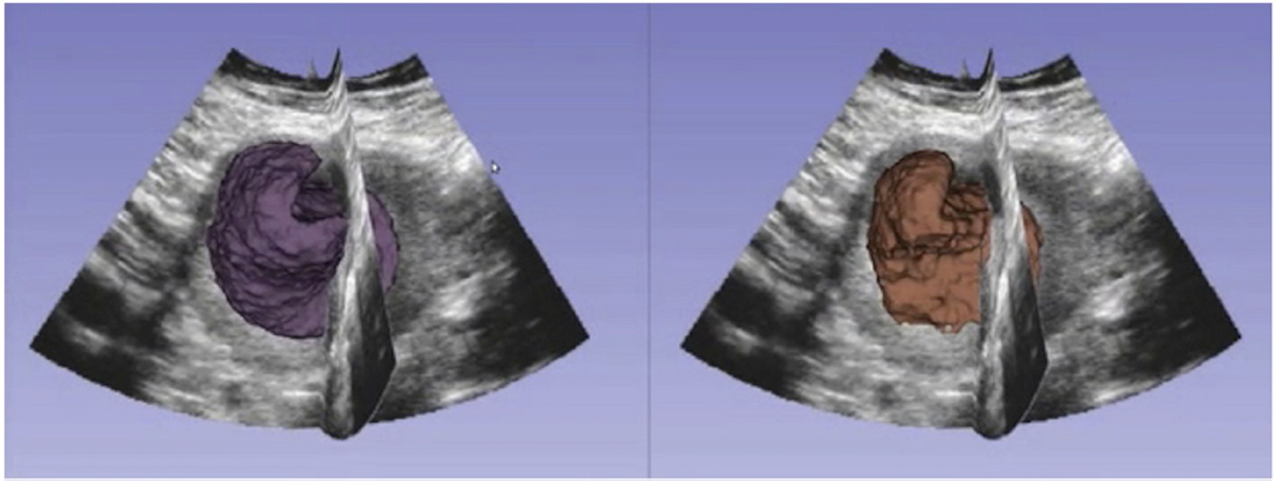
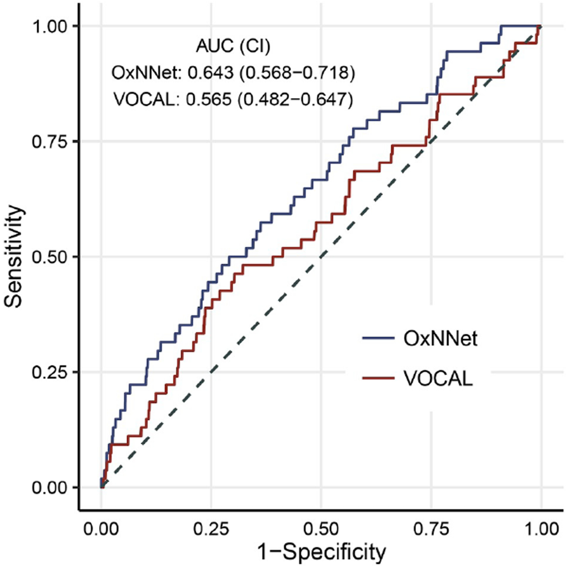
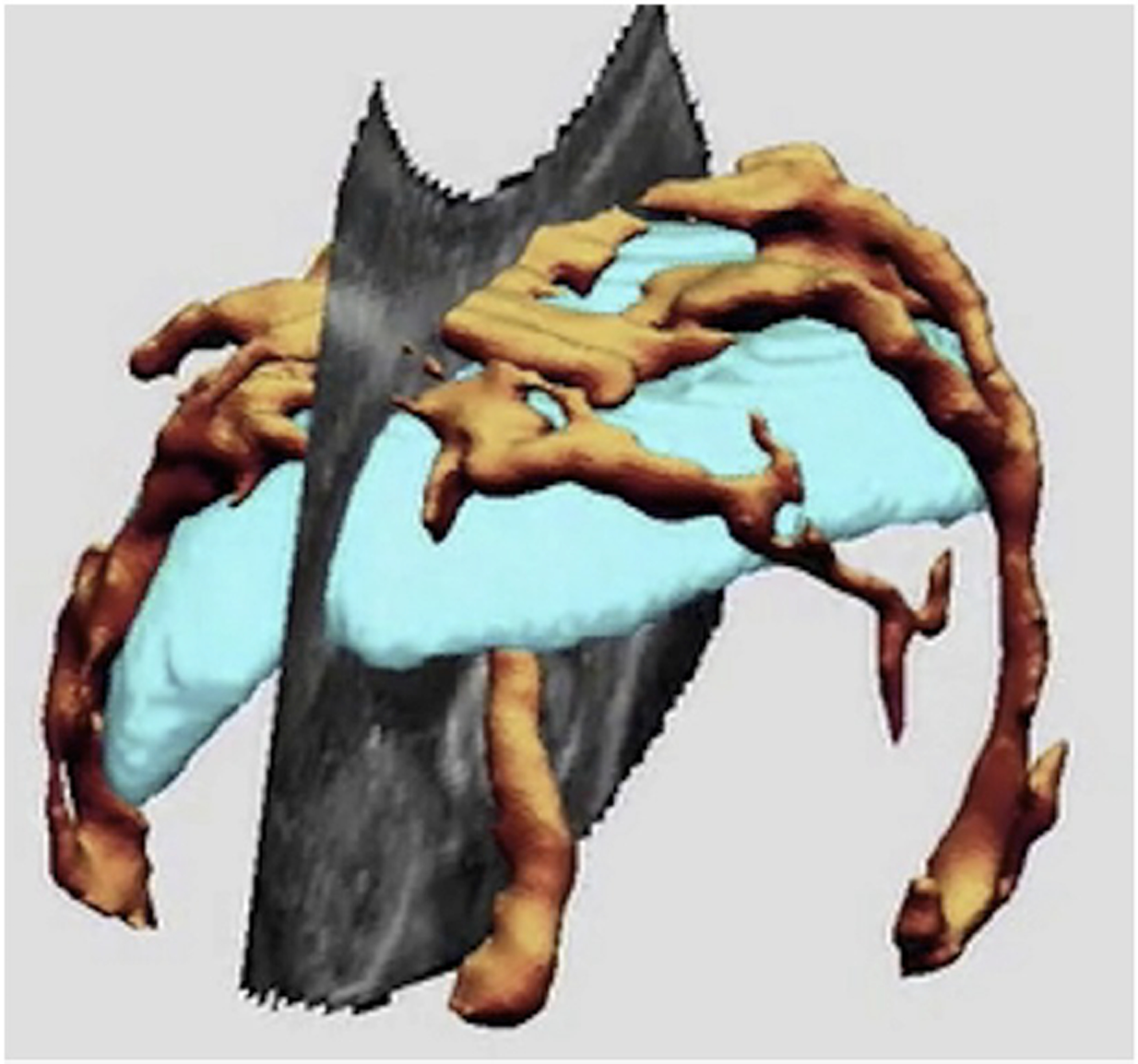
Fetal growth restriction (FGR) is a major cause of perinatal morbidity and mortality. Identifying which pregnancies are at risk of FGR facilitates enhanced surveillance and early delivery before fetal demise can ensue. However, existing risk stratification strategies yield an unacceptably low detection rate. A robust and reliable first trimester screening test for FGR would not only enable high-risk women to be appropriately monitored but would facilitate future trials for possible interventions to enhance fetal growth. Both the volume and vascularity of the first trimester placenta has been demonstrated to be linked to adverse pregnancy outcomes including FGR and pre-eclampsia. The investigation of novel ultrasound markers for FGR are discussed along with the development of methods for fully automatic placental volume estimation which has the potential for use as part of a multi-variable population-based screening test.
Keywords: Fetal growth restriction; Placental volume; Power Doppler; Screening; Stillbirth; Ultrasound.
Copyright © 2019 Elsevier Ltd. All rights reserved.
Conflict of interest statement
Declaration of interests
The authors have nothing to declare.
Figures
References
-
- Poon LC, et al. , Reference range of birth weight with gestation and first-trimester prediction of small-for-gestation neonates. Prenat Diagn, 2011. 31(1): p. 58–65. - PubMed
-
- Wen SW, et al. , Smoking, maternal age, fetal growth, and gestational age at delivery. Am J Obstet Gynecol, 1990. 162(1): p. 53–8. - PubMed
-
- Clausson B, Cnattingius S, and Axelsson O, Preterm and term births of small for gestational age infants: a population-based study of risk factors among nulliparous women. Br J Obstet Gynaecol, 1998. 105(9): p. 1011–7. - PubMed
-
- Salafia CM, Charles AK, and Maas EM, Placenta and fetal growth restriction. Clin Obstet Gynecol, 2006. 49(2): p. 236–56. - PubMed
Publication types
MeSH terms
Grants and funding
LinkOut - more resources
Full Text Sources
Miscellaneous
