

Interactive and Multifactorial Mechanisms of Calcific Vascular and Valvular Disease
- PMID: 31279666
- PMCID: PMC6708492
- DOI: 10.1016/j.tem.2019.06.001
Interactive and Multifactorial Mechanisms of Calcific Vascular and Valvular Disease
Abstract
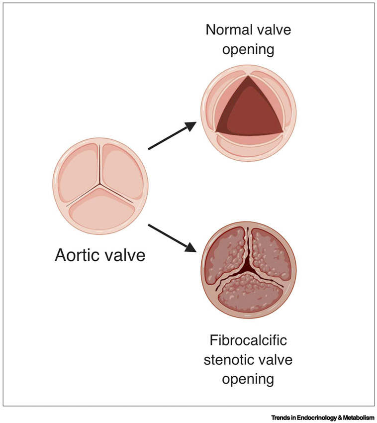
Calcific vascular and valvular disease (CVVD) is widespread and has major health consequences. Although coronary artery calcification has long been associated with hyperlipidemia and increased mortality, recent evidence suggests that its progression is increased in association with cholesterol-lowering HMG-CoA reductase inhibitors ('statins') and long-term, high-intensity exercise. A nationwide trial showed no cardiovascular benefit of vitamin D supplements. Controversy remains as to whether calcium deposits in plaque promote or prevent plaque rupture. CVVD appears to occur through mechanisms similar to those of intramembranous, endochondral, and osteophytic skeletal bone formation. New evidence implicates autotaxin, endothelial-mesenchymal transformation, and microRNA and long non-coding RNA (lncRNA) as novel regulatory factors. New therapeutic options are being developed.
Keywords: atherosclerosis; bone; calcification; cardiovascular; valvular disease.
Copyright © 2019 Elsevier Ltd. All rights reserved.
Figures
References
-
- Budoff MJ et al. (2007) Long-term prognosis associated with coronary calcification: observations from a registry of 25,253 patients. J Am Coll Cardiol 49 (18), 1860–70. - PubMed
-
- O'Rourke RA et al. (2000) American College of Cardiology/American Heart Association Expert Consensus document on electron-beam computed tomography for the diagnosis and prognosis of coronary artery disease. Circulation 102 (1), 126–40. - PubMed
