Review
. 2019 Apr:144:35-56.
doi: 10.1016/j.addr.2019.07.002. Epub 2019 Jul 4.

Integrating nanomedicine into clinical radiotherapy regimens

Affiliations
Review

Integrating nanomedicine into clinical radiotherapy regimens

Allison N DuRoss et al. Adv Drug Deliv Rev. 2019 Apr.

Abstract

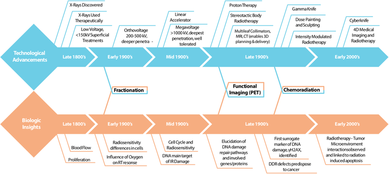
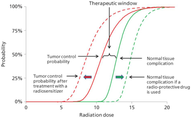
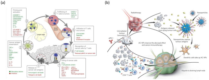
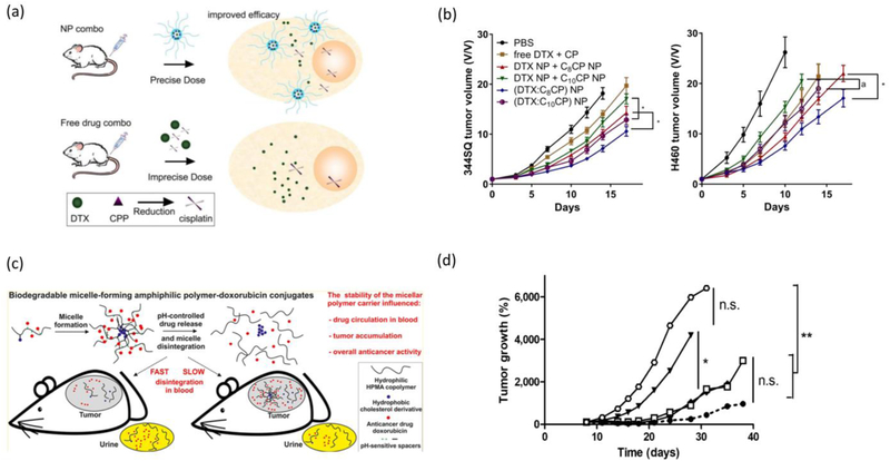
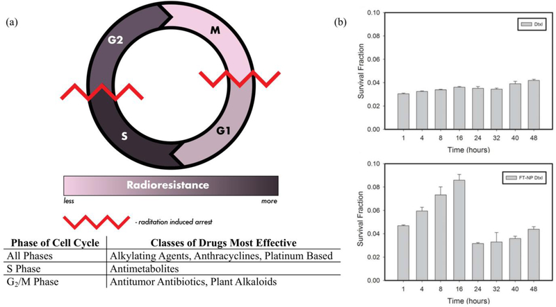
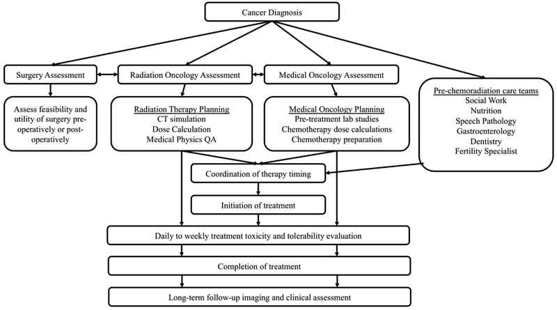
While the advancement of clinical radiotherapy was driven by technological innovations throughout the 20th century, continued improvement relies on rational combination therapies derived from biological insights. In this review, we highlight the importance of combination radiotherapy in the era of precision medicine. Specifically, we survey and summarize the areas of research where improved understanding in cancer biology will propel the field of radiotherapy forward by allowing integration of novel nanotechnology-based treatments.

Keywords: Cancer; Chemoradiotherapy; Dosing; Drug delivery; Multimodal therapy; Nanoparticles; Radiation therapy.


Figures

Figure 1.
Figure 1.
Timeline mapping seminal biological findings and technological advancements that have propelled radiation therapy to its current standard. Here we visualize the interplay between several insights and advancements, such as that of PET imaging, where an innovation has allowed physicians and scientists to elucidate biologic function thus improving combination therapy strategies.
Figure 2.
Figure 2.
The therapeutic window of radiation therapy is a balance between tumor control and normal tissue complications. Addition of radiosensitizers reduce the dose of radiation required for the same tumor control probability. Addition of radio-protective drugs increase the dose of radiation required for the same probability of normal tissue complications. Together, radiosensitizers and radio-protective drugs can enhance the therapeutic window of RT. Reprinted with permission from [19].
Figure 3.
Figure 3.
Schematic illustration of the types of nanoparticles discussed in this review and the benefits of utilizing these carriers to delivery poorly soluble or toxic therapeutic agents.
Figure 4.
Figure 4.
(a) Quantification of cleaved-caspase 3 (CC3) staining in tumor sections of mice treated with various nanoparticles with or without IR compared to no treatment control. Adapted with permission from [111]. (b) Tumor sections stained with CC3 for (i) empty micelle, (ii) dual-loaded micelle, (iii) empty micelle + IR, and (iv) dual-loaded micelle + IR. Inset sections represent the corresponding boxed region in whole-tumor image. Outlined sections are designed to illustrate the extent of CC3 staining and thus apoptosis. Adapted with permission from [112].
Figure 5.
Figure 5.
Radiation influences the tumor microenvironment in many ways and recently research has begun to further understand these effects and the complex way they may be exploited to enhanced tumoral accumulation of nanoparticles. (a) Graphical abstract highlights some of these influences. Adapted with permission from [135]. (b) (top) Pretreatment with radiation decreased IFP 24 h post-treatment and the intratumoral bulk accumulation in MDA-MB-231 tumors. (bottom) Tumor IFP was low (4.5 ± 1.5 mmHg) but not negligible in the 4T1 tumors. A significant change in IFP following radiation was not observed and radiation does not improve the bulk accumulation, intratumoral distribution. In in vivo studies radiation or followed by Doxil resulted in a modest increase in tumor growth delay compared to all other treatments in the 4T1 tumor model. Adapted with permission from [143].
Figure 6.
Figure 6.
Radiation influences on tumoral nanoparticle accumulation. (a) Tumor accumulation of NIR-fluorescent polymeric micelles administered either 24 hours or 72 hours after the last dose of RT (3 consecutive daily doses of 5 Gy) in a 4T1 model. Adapted with permission from [112]. (b) Tumor accumulation of 3 different fluorescent nanoparticles administered 72 hours after a single dose of 5 Gy in 4T1 bearing mice. Adapted with permission from [146].
Figure 7.
Figure 7.
P-selectin targeting for enhancing RT (a) synthetic scheme of the preparation of fucoidan-encapsulated paclitaxel nanoparticles (FiPAX). (b) Fluorescence images of human endothelial monolayer treated with TNF-alpha and IR (6 Gy) to induce P-selectin. Red, NIR dye in FiPAX or control DexPAX nanoparticles; green, CellMask membrane stain; blue, DAPI nuclear stain. Scale bar, 5 μm. (c) Immunofluorescence measurements of 3LL tumors extracted from mice after radiation treatment. Green, P-selectin; blue, DAPI nuclear stain. Scale bar, 50 μm. (d) Tumor growth inhibition after irradiation and single-dose administration of drug treatments. All treatments were given on day 10 after tumor inoculation. Adapted with permission from [155].
Figure 8.
Figure 8.
Immunotherapy and RT (a) cancer-immunity cycle with stimulatory and inhibitory factors. Stimulatory factors shown in green promote immunity, whereas inhibitors shown in red help keep the process in check and reduce immune activity and/or prevent autoimmunity. Immune checkpoint proteins, such as CTLA4, can inhibit the development of an active immune response by acting primarily at the level of T cell development and proliferation (step 3). We distinguish these from immune rheostat (“immunostat”) factors, such as PD-L1, can have an inhibitory function that primarily acts to modulate active immune responses in the tumor bed (step 7). Examples of such factors and the primary steps at which they can act are shown. Although not illustrated, it is important to note that intratumoral T regulatory cells, macrophages, and myeloid-derived suppressor cells are key sources of many of these inhibitory factors. Reproduced with permission from [157]. (b) Schematic illustration depicting the utilization of antigen-capturing nanoparticles (AC-NPs) to improve cancer immunotherapy. After RT, ACNPs will bind to tumor antigens and improve their presentation to dendritic cells. Reproduced with permission from [159].
Figure 9.
Figure 9.
Influence of drug combinations on in vivo efficacy (a) nanoparticle co-encapsulation of docetaxel and cisplatin allows for accurate exposure of the tumor site to both drugs. Comparatively, free drug dosing may lead to variations in tumor drug exposure, reducing potency. (b) Free drug and nanoparticle formulation in vivo efficacy in 344SQ and H460 murine xenograft models. Mice were treated with combinations of either the free drugs, singly loaded nanoparticles, or dually loaded nanoparticles. Tumor microenvironment-responsive nanoparticles. Reproduced with permission from [164]. (c) pH responsive nanoparticle of varying stability influence overall anticancer activity. (d) tumor growth and resistance in murine EL4 T-cell lymphoma treated with micellar doxorubicin conjugates. Reproduced with permission from [165].
Figure 10.
Figure 10.
(a) Radioresistance and the cell cycle. Cellular radioresistance varies depending on the stage of cell cycle with mitosis being the least radioresistant and synthesis being the most radioresistant. Due to their mechanism of action, classes of radiosensitizers are more effective at different cell cycle phases, demonstrated here. Red jagged lines depict cell cycle checkpoints that are triggered when DNA is damaged by radiation. (b) Timing plays an important role in the optimal combination of RT and nanoparticle delivered chemotherapeutics. KB cells treated with docetaxel, a plant alkaloid which induces G2/M phase arrest, (Dxtl) (top) or FT-NP Dtxl (bottom) irradiated with 4 Gy at the indicated times and evaluated for clonogenicity. Adapted with permission from [15].
Figure 11.
Figure 11.
A detailed flow chart detailing the intricate treatment plan that underlies each therapeutic regimen in oncology. Interdisciplinary collaboration between a multitude of medical teams is necessary for optimal patient experiences and outcomes.

Similar articles

Cited by

References

    1. Siegel RL, Miller KD, Jemal A, Cancer statistics, 2019, CA. Cancer J. Clin 69 (2019) 7–34. - PubMed
    1. Miller KD, Siegel RL, Lin CC, Mariotto AB, Kramer JL, Rowland JH, Stein KD, Alteri R, Jemal A, Cancer treatment and survivorship statistics, 2016, CA. Cancer J. Clin 66 (2016) 271–289. - PubMed
    1. Palacios Eito A, Cabezas SG, Ugalde PF, del Campo ER, Romero AO, del M Martín MP, Arjona JMR, Paredes MM, Characterization and adequacy of the use of radiotherapy and its trend in time, Radiother. Oncol 106 (2013) 260–265. - PubMed
    1. Delaney G, Jacob S, Featherstone C, Barton M, The role of radiotherapy in cancer treatment, Cancer. 104 (2005) 1129–1137. - PubMed
    1. Barcellos-Hoff MH, Park C, Wright EG, Radiation and the microenvironment – tumorigenesis and therapy, Nat. Rev. Cancer 5 (2005) 867–875. - PubMed

Publication types