. 2019 Dec;56(12):8589-8602.
doi: 10.1007/s12035-019-01691-w. Epub 2019 Jul 6.

Deletion of Arginase 2 Ameliorates Retinal Neurodegeneration in a Mouse Model of Multiple Sclerosis

Affiliations

Deletion of Arginase 2 Ameliorates Retinal Neurodegeneration in a Mouse Model of Multiple Sclerosis

Chithra D Palani et al. Mol Neurobiol. 2019 Dec.

Abstract

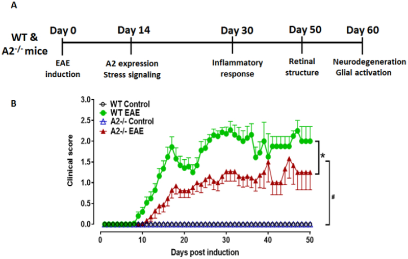
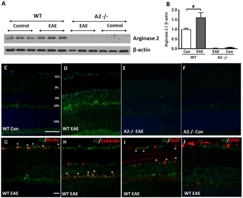
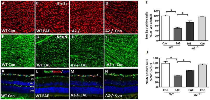
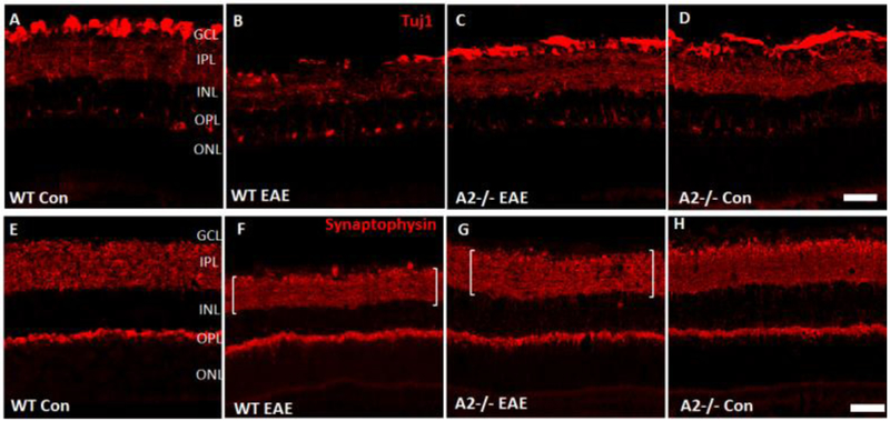
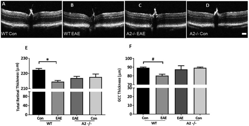
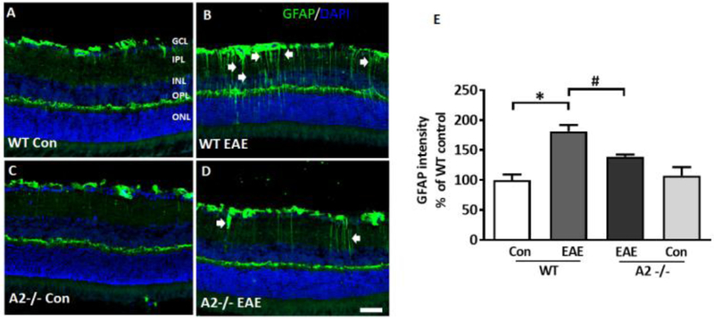
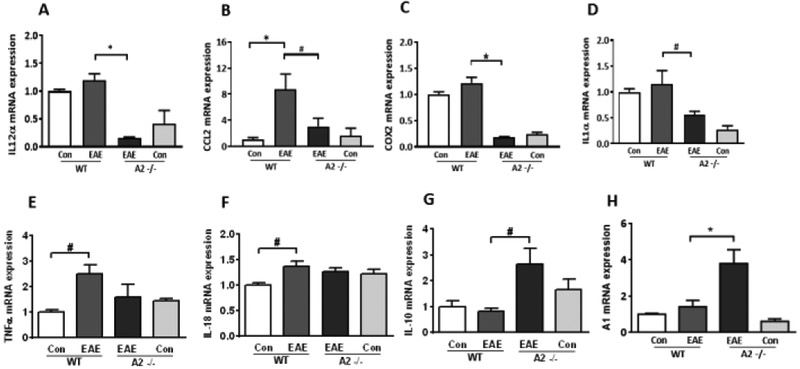
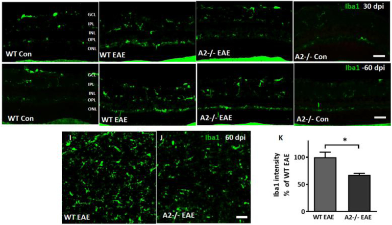
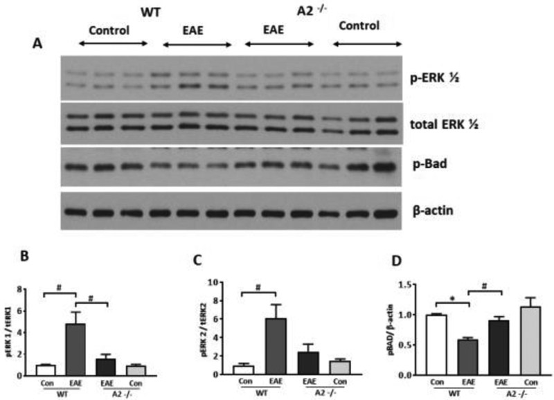
Optic neuritis is a major clinical feature of multiple sclerosis (MS) and can lead to temporary or permanent vision loss. Previous studies from our laboratory have demonstrated the critical involvement of arginase 2 (A2) in retinal neurodegeneration in models of ischemic retinopathy. The current study was undertaken to investigate the role of A2 in MS-mediated retinal neuronal damage and degeneration. Experimental autoimmune encephalomyelitis (EAE) was induced in wild-type (WT) and A2 knockout (A2-/-) mice. EAE-induced motor deficits, loss of retinal ganglion cells, retinal thinning, inflammatory signaling, and glial activation were studied in EAE-treated WT and A2-/- mice and their respective controls. Increased expression of A2 was observed in WT retinas in response to EAE induction. EAE-induced motor deficits were markedly reduced in A2-/- mice compared with WT controls. Retinal flat mount studies demonstrated a significant reduction in the number of RGCs in WT EAE retinas in comparison with normal control mice. A significant improvement in neuronal survival was evident in retinas of EAE-induced A2-/- mice compared with WT. RNA levels of the proinflammatory molecules CCL2, COX2, IL-1α, and IL-12α were significantly reduced in the A2-/- EAE retinas compared with WT EAE. EAE-induced activation of glia (microglia and Müller cells) was markedly reduced in A2-/- retinas compared with WT. Western blot analyses showed increased levels of phospho-ERK1/2 and reduced levels of phospho-BAD in the WT EAE retina, while these changes were prevented in A2-/- mice. In conclusion, our studies establish EAE as an excellent model to study MS-mediated retinal neuronal damage and suggest the potential value of targeting A2 as a therapy to prevent MS-mediated retinal neuronal injury.

Keywords: Arginase 2; EAE; Neurodegeneration; Optic neuritis; Retina; Retinal ganglion cells.


Conflict of interest statement

Conflict of Interest:

The authors declare no conflict of interest.

Figures

Figure 1:
Figure 1:. EAE induction and evaluation of clinical scores.
A) Schematic representation of the time points of EAE induction and experiments performed. B) Animals were evaluated every day and the clinical scores were recorded according to a 0–5 scale (see methods section for details). WT EAE mice showed progressively increasing clinical score starting at day 9. A2−/− EAE mice showed significantly lower clinical score as compared to WT EAE mice. Both control groups showed no signs of motor deficits. *p<0.05, A2−/− EAE vs WT EAE for days 11-21, 24-37, 41-43 and 47. #p<0.05, WT and A2−/− EAE groups vs respective control groups starting at days 11 and 14 respectively, and till the end of the experiment. N = 17-26 per group. Four animals in WT EAE and two in A2 −/− EAE group died or were sacrificed due to weight loss. Data are presented as mean ± SEM.
Figure 2:
Figure 2:. Expression of arginase 2 is increased in the WT EAE retina.
A, B) Western blotting and quantification showed a significant increase in A2 expression in the WT EAE retinas at 14-day post immunization as compared to WT control. C-F) Representative immunofluorescence images showing increased expression of A2 in WT EAE retina as compared to WT control. No expression of A2 is observed in A2−/− retinas. G-J) Colocalization studies showing expression of A2 in retinal ganglion cells (Brn3a), amacrine cells (ChAT), and horizontal cells (calbindin). * indicate areas of colocalization. Scale bar: 50 μm. Data are presented as mean ± SEM #p< 0.05. N = 4-5.
Figure 3:
Figure 3:. A2 deletion protects against EAE-induced RGC loss.
Representative confocal images showing the immunolabeling of retinal flat mounts with Brn3a (A-D) and NeuN in the GCL layer (F-I). Quantitative analysis demonstrating significant loss of NeuN-positive (E) and Brn3a positive (J) cells in the GCL in response to EAE induction. A2 deletion significantly protected against the EAE induced neuronal loss. Data are presented as mean ± SEM. * (P < 0.01); # (p< 0.05). N = 6-7 per group. Scale bar: 50 μm. K-N) Confocal images of retinal cryostat sections immunostained with NeuN, Brn3a and DAPI. Arrows indicate areas of cell loss. Scale bar: 50 μm. GCL, ganglion cell layer; INL, inner nuclear layer; IPL, inner plexiform layer; ONL, outer nuclear layer; OPL, outer plexiform layer. N= 4-6, and representative images are presented.
Fig 4.
Fig 4.. EAE-induced neurodegeneration in the inner retina is decreased by A2 deletion.
Confocal images of retinal cryostat sections showing the immunolabeling of Tuj1 (A-D), a marker for RGCs and their axons, and synaptophysin (E-H), a pre-synaptic marker. A marked reduction is observed in the expression of both Tuj1 and synaptophysin in WT EAE retina, while A2 deletion markedly improved their levels. EAE-induced downregulation of both markers was markedly improved in A2 retinas. Brackets indicate synaptic zone thickness. Scale bar: 100 μm. GCL, ganglion cell layer; INL, inner nuclear layer; IPL, inner plexiform layer; ONL, outer nuclear layer; OPL, outer plexiform layer. N= 4-6, and representative images are presented.
Figure 5:
Figure 5:. EAE induced retinal thinning is abrogated by A2 deletion.
A-D) Representative B-scan images from control and EAE retinas, using Spectral Domain-Optical Coherence Tomography (SD-OCT). Scale bar: 100 μm E-F) Quantitative analysis showing significant reduction in the thickness of total retina and IPL (inner plexiform layer) in WT EAE compared to WT controls. These changes are partially rescued in the retinas of the A2−/− mice but the differences are not statistically significant. Data are presented as mean ± SEM. *p<0.01; #p< 0.05. N= 8-13.
Figure 6:
Figure 6:. A2 deletion ameliorates EAE-induced glial activation.
A-D) Immunofluorescence staining of retinal sections (60 days post immunization) using GFAP antibody, demonstrating activation of glial cells in the WT EAE retina. Deletion of A2 markedly reduced this effect. Arrows indicate activated Muller cell processes. (E) Quantification of the GFAP immunofluorescence intensity (from nerve fiber layer to outer limiting membrane) showing the increased GFAP level in the WT EAE retinal sections. A2 deletion significantly reduced this effect. N =4-6, and representative images are presented. GCL, ganglion cell layer; IPL, inner plexiform layer; INL, inner nuclear layer; OPL, outer plexiform layer; ONL, outer nuclear layer. Scale bar: 50 μm. Data are presented as mean ± SEM. *p<0.01; #p<0.05.
Figure 7:
Figure 7:. A2 deletion suppresses EAE-induced inflammatory response.
A-D) RT-PCR analysis demonstrating changes in mRNA levels of pro-inflammatory cytokines and chemokines in EAE retinas (30 days post immunization). IL-12α (A), CCL2 (B), COX2 (C) and IL-1α (D) mRNA levels were markedly reduced by A2 deletion in EAE groups. E-F) EAE-induced increases in TNFα and IL-18 were not affected by A2 deletion. G-H) A2 deletion significantly increased the anti-inflammatory molecules IL-10 and A1 as compared to WT EAE and control groups. Data are presented as mean ± SEM. *p<0.01; #p< 0.05. N = 3-10.
Figure 8:
Figure 8:. A2 deletion ameliorates EAE-induced microglial activation.
A-D) Immunofluorescence staining for Iba1 (a microglia/macrophage marker) on retinal sections (30 days post immunization (dpi)) showed increased microglial activation in the WT EAE retina. A2−/− EAE retina showed reduced microglial activation. EAE-induced microglial activation continued to be observed in WT EAE retina at 60 days post immunization (dpi), A2 deletion reduced this activation (E-H). N =4-6, and representative images are presented. GCL, ganglion cell layer; IPL, inner plexiform layer; INL, inner nuclear layer; OPL, outer plexiform layer; ONL, outer nuclear layer. Scale bar: 50 μm. I-J) Iba1 immunostaining on retinal flatmounts from WT EAE and A2−/− EAE retinas. K) Quantification of Iba1 fluorescence intensity on retinal flat mounts showing significant downregulation in the A2−/− EAE group as compared to WT EAE. Data are presented as mean ± SEM. *P <0.01. Scale bar:50 μm.
Figure 9:
Figure 9:. Deletion of A2 reduced EAE induced stress signaling in the retina.
A) Western blot studies showing increased levels of p-ERK1/2 and reduced p-BAD levels in WT EAE retina. These changes were reversed in A2−/− EAE retina. B-D) Quantitative analysis of western blots demonstrate significantly increased levels of p-ERK1/2 and reduced levels of p-BAD in the WT EAE retina compared to WT control. Deletion of A2 altered the EAE-induced increase in p-ERK1 levels and reduction in p-BAD levels. Data are presented as mean ± SEM. *P < 0.01; #p< 0.05. N=3-6.

References

    1. Balcer LJ, Miller DH, Reingold SC, Cohen JA. Vision and vision-related outcome measures in multiple sclerosis. Brain. 2015;138:11–27 - PMC - PubMed
    1. Sakai RE, Feller DJ, Galetta KM, Galetta SL, Balcer LJ. Vision in multiple sclerosis: The story, structure-function correlations, and models for neuroprotection. J Neuroophthalmol. 2011;31:362–373 - PMC - PubMed
    1. Galetta SL, Villoslada P, Levin N, Shindler K, Ishikawa H, Parr E, et al. Acute optic neuritis: Unmet clinical needs and model for new therapies. Neurology(R) neuroimmunology & neuroinflammation. 2015;2:e135. - PMC - PubMed
    1. Sakai T, Ishihara T, Higaki M, Akiyama G, Tsuneoka H. Therapeutic effect of stealth-type polymeric nanoparticles with encapsulated betamethasone phosphate on experimental autoimmune uveoretinitis. Investigative ophthalmology & visual science. 2011;52:1516–1521 - PubMed
    1. Walter SD, Ishikawa H, Galetta KM, Sakai RE, Feller DJ, Henderson SB, et al. Ganglion cell loss in relation to visual disability in multiple sclerosis. Ophthalmology. 2012;119:1250–1257 - PMC - PubMed

MeSH terms