

Prospective non-invasive evaluation of CXCR4 expression for the diagnosis of MALT lymphoma using [68Ga]Ga-Pentixafor-PET/MRI
- PMID: 31281504
- PMCID: PMC6587159
- DOI: 10.7150/thno.31032
Prospective non-invasive evaluation of CXCR4 expression for the diagnosis of MALT lymphoma using [68Ga]Ga-Pentixafor-PET/MRI
Abstract
MALT lymphomas express the chemokine receptor CXCR4 on a regular basis, and [68Ga]Ga-Pentixafor-PET has been shown to quantify CXCR4 expression non-invasively. We, therefore, aimed to evaluate [68Ga]Ga-Pentixafor-PET/MRI for the non-invasive assessment of MALT lymphomas.
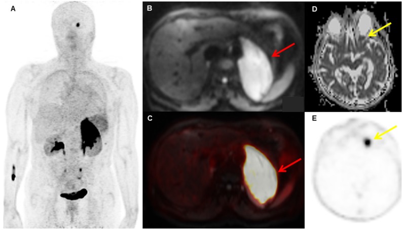
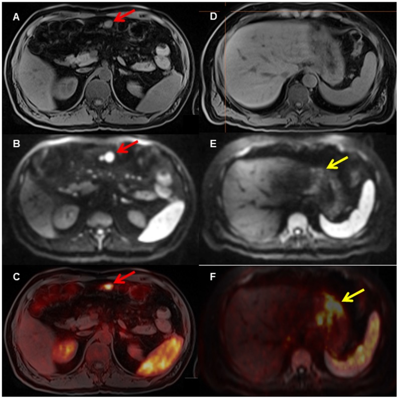
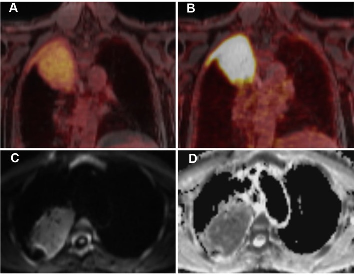
Methods: We included 36 MALT lymphoma patients, who had not undergone previous systemic or radiation therapy, in our prospective, IRB-approved, proof-of-concept study. Involved anatomic regions were the orbit (n=14), stomach (n=10), lungs (n=5), and other sites (soft-tissues n=3; adrenal gland, tonsils, parotid gland, and urinary bladder n=1, respectively). MRI sequences included an axial 2-point Dixon T1 VIBE SPAIR 3D sequence for PET attenuation correction; a coronal T2 HASTE sequence; and an axial echo-planar imaging SPAIR-based diffusion-weighted sequence (DWI) obtained during free-breathing (b-values, 50 and 800), with corresponding ADC (apparent diffusion coefficient) maps.
Results: In 33/36 patients, there were MALT lymphomas with an increased uptake of [68Ga]Ga-Pentixafor; all current lymphoma manifestations showed an increased uptake and, accordingly, were positive on the PET/MRI. The remaining three patients had undergone surgery for their orbital MALT lymphomas prior to PET/MRI. Mean SUVmax was 8.6 ± 4.7, mean SUVmean was 4.7 ± 1.8, and mean SUVpeak was 8.0 ± 4.2. The mean SUVmax of the liver was 1.8, and the mean tumor-to-liver ratio was 2.9 ± 2.0. There were no significant differences in SUVmax (P=0.22), SUVmean (P=0.53), SUVpeak (P=0.29), or SUVt/l (P=0.92) between the four anatomic regions (orbit, stomach, lungs, other). The mean tumor volume was 146 ± 499.
Conclusions: Our results thus indicate that [68Ga]Ga-Pentixafor-PET is feasible for the assessment of MALT lymphomas, with a good tumor-to-background ratio in terms of radiotracer uptake.
Keywords: CXCR4; MALT lymphoma; PET; PET/MRI; [68Ga]-Pentixafor.
Conflict of interest statement
Competing Interests: H.W. is a founder and shareholder of Scintomics. S.K. is CEO of Scintomics. M.Ma. received research grants and honoraria for presentations from Siemens Healthineers. A.R.H. received research grants from Siemens Healthineers. The other authors declare no competing financial interests.
Figures
Similar articles
-
CXCR4 PET/MRI for follow-up of gastric mucosa-associated lymphoid tissue lymphoma after first-line Helicobacter pylori eradication.Blood. 2022 Jan 13;139(2):240-244. doi: 10.1182/blood.2021013239. Blood. 2022. PMID: 34525196 Free PMC article.
-
[68Ga]Pentixafor PET/MR imaging of chemokine receptor 4 expression in the human carotid artery.Eur J Nucl Med Mol Imaging. 2019 Jul;46(8):1616-1625. doi: 10.1007/s00259-019-04322-7. Epub 2019 Apr 19. Eur J Nucl Med Mol Imaging. 2019. PMID: 31004184 Free PMC article.
-
[68Ga]Ga-Pentixafor PET/MRI for CXCR4 Imaging of Chronic Lymphocytic Leukemia: Preliminary Results.Invest Radiol. 2018 Jul;53(7):403-408. doi: 10.1097/RLI.0000000000000469. Invest Radiol. 2018. PMID: 29642081
-
Positron emission tomography/magnetic resonance imaging (PET/MRI) vs. gastroscopy: Can it improve detection of extranodal marginal zone lymphomas of the stomach following H. pylori treatment?Expert Rev Hematol. 2022 Jul;15(7):565-571. doi: 10.1080/17474086.2022.2089110. Epub 2022 Jun 21. Expert Rev Hematol. 2022. PMID: 35695746 Free PMC article. Review.
-
Advances in PET Imaging of the CXCR4 Receptor: [68Ga]Ga-PentixaFor.Semin Nucl Med. 2024 Jan;54(1):163-170. doi: 10.1053/j.semnuclmed.2023.09.002. Epub 2023 Nov 3. Semin Nucl Med. 2024. PMID: 37923671 Free PMC article. Review.
Cited by
-
[68Ga]Ga-Pentixafor and Sodium [18F]Fluoride PET Can Non-Invasively Identify and Monitor the Dynamics of Orthodontic Tooth Movement in Mouse Model.Cells. 2022 Sep 21;11(19):2949. doi: 10.3390/cells11192949. Cells. 2022. PMID: 36230911 Free PMC article.
-
Preliminary evidence of imaging of chemokine receptor-4-targeted PET/CT with [68Ga]pentixafor in non-Hodgkin lymphoma: comparison to [18F]FDG.EJNMMI Res. 2020 Aug 5;10(1):89. doi: 10.1186/s13550-020-00681-7. EJNMMI Res. 2020. PMID: 32757068 Free PMC article.
-
Current Role of Functional Imaging in the Management of Lymphoma.Curr Oncol Rep. 2021 Nov 4;23(12):144. doi: 10.1007/s11912-021-01127-6. Curr Oncol Rep. 2021. PMID: 34735647 Review.
-
Research Progress of CXCR4-Targeting Radioligands for Oncologic Imaging.Korean J Radiol. 2023 Sep;24(9):871-889. doi: 10.3348/kjr.2023.0091. Korean J Radiol. 2023. PMID: 37634642 Free PMC article. Review.
-
What you always wanted to know about gastric MALT-lymphoma: a focus on recent developments.Ther Adv Med Oncol. 2021 Jul 23;13:17588359211033825. doi: 10.1177/17588359211033825. eCollection 2021. Ther Adv Med Oncol. 2021. PMID: 34621332 Free PMC article. Review.
References
-
- Zucca E, C Copie-Bergman U Ricardi, C Thieblemont M Raderer, M Ladetto et al. Gastric marginal zone lymphoma of MALT type: ESMO Clinical Practice Guidelines for diagnosis, treatment and follow-up. Ann Oncol. 2013;24(Suppl 6):144–8. - PubMed
-
- Mayerhoefer ME, G Karanikas K Kletter, H Prosch B Kiesewetter, C Skrabs et al. Evaluation of diffusion-weighted MRI for pretherapeutic assessment and staging of lymphoma: results of a prospective study in 140 patients. Clin Cancer Res. 2014;20:2984–93. - PubMed
-
- Ruskone-Fourmestraux A, W Fischbach BM Aleman, H Boot MQ Du, F Megraud et al. EGILS consensus report. Gastric extranodal marginal zone B-cell lymphoma of MALT. Gut. 2011;60:747–58. - PubMed
-
- Raderer M, S Wohrer B Streubel, M Troch K Turetschek, U Jager et al. Assessment of disease dissemination in gastric compared with extragastric mucosa-associated lymphoid tissue lymphoma using extensive staging: a single-center experience. J Clin Oncol. 2006;24:3136–41. - PubMed
-
- Dreyling M, C Thieblemont A Gallamini, L Arcaini E Campo, O Hermine et al. ESMO Consensus conferences: guidelines on malignant lymphoma. part 2: marginal zone lymphoma, mantle cell lymphoma, peripheral T-cell lymphoma. Ann Oncol. 2013;24:857–77. - PubMed
MeSH terms
Substances
Grants and funding
LinkOut - more resources
Full Text Sources
