

Chemiluminescence and Bioluminescence Imaging for Biosensing and Therapy: In Vitro and In Vivo Perspectives
- PMID: 31281531
- PMCID: PMC6592176
- DOI: 10.7150/thno.33228
Chemiluminescence and Bioluminescence Imaging for Biosensing and Therapy: In Vitro and In Vivo Perspectives
Abstract
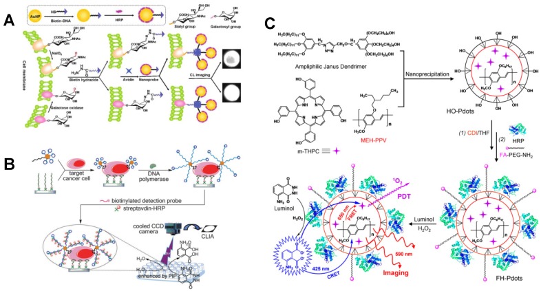
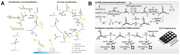
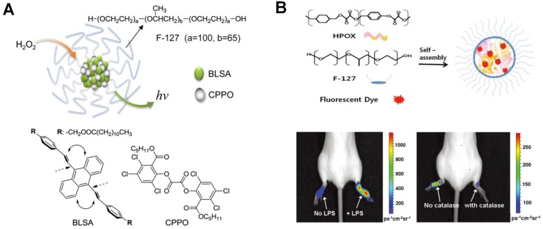
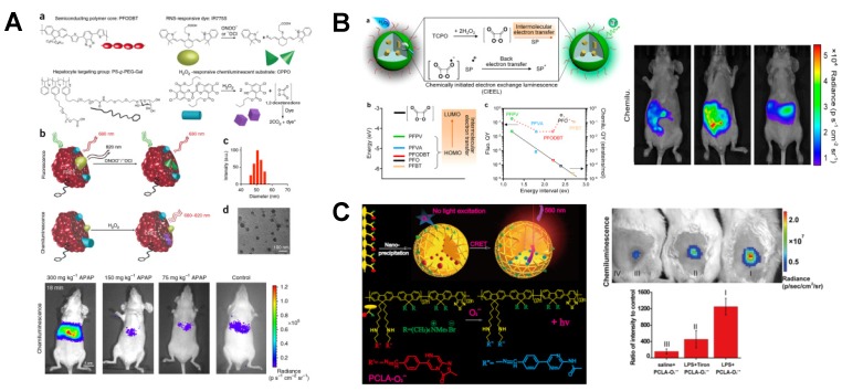
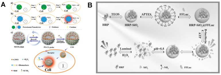
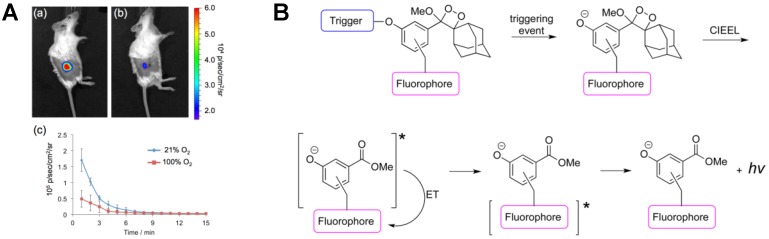
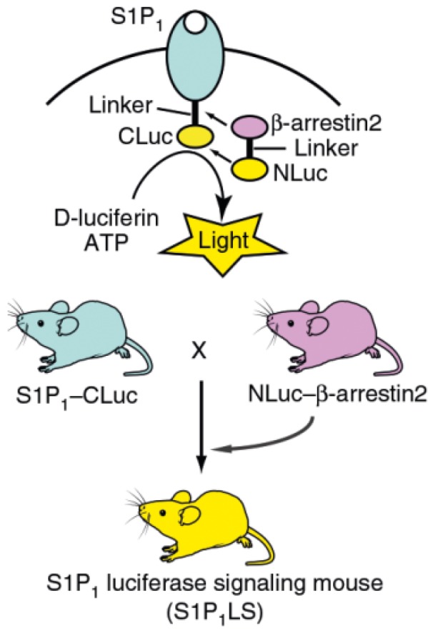
Chemiluminescence (CL) and bioluminescence (BL) imaging technologies, which require no external light source so as to avoid the photobleaching, background interference and autoluminescence, have become powerful tools in biochemical analysis and biomedical science with the development of advanced imaging equipment. CL imaging technology has been widely applied to high-throughput detection of a variety of analytes because of its high sensitivity, high efficiency and high signal-to-noise ratio (SNR). Using luciferase and fluorescent proteins as reporters, various BL imaging systems have been developed innovatively for real-time monitoring of diverse molecules in vivo based on the reaction between luciferin and the substrate. Meanwhile, the kinetics of protein interactions even in deep tissues has been studied by BL imaging. In this review, we summarize in vitro and in vivo applications of CL and BL imaging for biosensing and therapy. We first focus on in vitro CL imaging from the view of improving the sensitivity. Then, in vivo CL applications in cells and tissues based on different CL systems are demonstrated. Subsequently, the recent in vitro and in vivo applications of BL imaging are summarized. Finally, we provide the insight into the development trends and future perspectives of CL and BL imaging technologies.
Keywords: bioimaging; bioluminescence; biomedicine; biosensing; chemiluminescence.
Conflict of interest statement
Competing Interests: The authors have declared that no competing interest exists.
Figures
Similar articles
-
Recent Applications and Future Perspectives of Chemiluminescent and Bioluminescent Imaging Technologies.Chem Biomed Imaging. 2023 Apr 13;1(4):297-314. doi: 10.1021/cbmi.2c00002. eCollection 2023 Jul 24. Chem Biomed Imaging. 2023. PMID: 39473938 Free PMC article. Review.
-
Analytical chemiluminescence and bioluminescence: latest achievements and new horizons.Anal Bioanal Chem. 2012 Jan;402(1):69-76. doi: 10.1007/s00216-011-5455-8. Epub 2011 Oct 16. Anal Bioanal Chem. 2012. PMID: 22002591 Review.
-
Development and Applications of Bioluminescent and Chemiluminescent Reporters and Biosensors.Annu Rev Anal Chem (Palo Alto Calif). 2019 Jun 12;12(1):129-150. doi: 10.1146/annurev-anchem-061318-115027. Epub 2019 Feb 20. Annu Rev Anal Chem (Palo Alto Calif). 2019. PMID: 30786216 Free PMC article. Review.
-
Steady-state bioluminescence of bacterial luciferase using electrochemical regeneration of flavin substrate and its application to inhibitory analysis.Bioelectrochemistry. 2009 Apr;75(1):67-70. doi: 10.1016/j.bioelechem.2008.12.002. Epub 2008 Dec 25. Bioelectrochemistry. 2009. PMID: 19162563
-
Recent progress in biomedical applications of persistent luminescence nanoparticles.Nanoscale. 2017 May 18;9(19):6204-6218. doi: 10.1039/c7nr01488k. Nanoscale. 2017. PMID: 28466913 Review.
Cited by
-
Gaussia Luciferase as a Reporter for Quorum Sensing in Staphylococcus aureus.Sensors (Basel). 2020 Aug 1;20(15):4305. doi: 10.3390/s20154305. Sensors (Basel). 2020. PMID: 32752273 Free PMC article.
-
The Cutting Edge of Disease Modeling: Synergy of Induced Pluripotent Stem Cell Technology and Genetically Encoded Biosensors.Biomedicines. 2021 Aug 5;9(8):960. doi: 10.3390/biomedicines9080960. Biomedicines. 2021. PMID: 34440164 Free PMC article. Review.
-
Self-Illuminating Agents for Deep-Tissue Optical Imaging.Front Bioeng Biotechnol. 2019 Nov 12;7:326. doi: 10.3389/fbioe.2019.00326. eCollection 2019. Front Bioeng Biotechnol. 2019. PMID: 31799247 Free PMC article. Review.
-
Small-Molecules as Chemiluminescent Probes to Detect Lipase Activity.Int J Mol Sci. 2022 Aug 12;23(16):9039. doi: 10.3390/ijms23169039. Int J Mol Sci. 2022. PMID: 36012304 Free PMC article.
-
Recombinase Polymerase Amplification-Based Biosensors for Rapid Zoonoses Screening.Int J Nanomedicine. 2023 Nov 6;18:6311-6331. doi: 10.2147/IJN.S434197. eCollection 2023. Int J Nanomedicine. 2023. PMID: 37954459 Free PMC article. Review.
References
-
- Aboul-Enein HY, Stefan R-I, van Staden JF, Zhang XR, Garcia-Campana AM, Baeyens WRG. Recent developments and applications of chemiluminescence sensors. Crit Rev Anal Chem. 2000;30:271–89.
-
- Roda A, Guardigli M, Pasini P, Mirasoli M, Michelini E, Musiani M. Bio- and chemiluminescence imaging in analytical chemistry. Anal Chim Acta. 2005;541:25–36.
-
- Jansen EHJM, Buskens CAF, van den Berg RH. A sensitive CCD image system for detection of chemiluminescent reactions. J Biolumin Chemilumin. 1989;3:53–7. - PubMed
-
- Créton R, Jaffe LF. Chemiluminescence microscopy as a tool in biomedical research. Biotechniques. 2001;31:1098–100. - PubMed
-
- Momeni N, Ramanathan K, Larsson PO, Danielsson B, Bengmark S, Khayyami M. CCD-camera based capillary chemiluminescent detection of retinol binding protein. Anal Chim Acta. 1999;387:21–7.
Publication types
MeSH terms
LinkOut - more resources
Full Text Sources
Other Literature Sources
