

Molecular Imaging in Ischemic Heart Disease
- PMID: 31281564
- PMCID: PMC6557873
- DOI: 10.1007/s12410-019-9500-x
Molecular Imaging in Ischemic Heart Disease
Abstract
Purpose of review: The purpose of this paper is to review current and new modalities to image key biological processes in ischemic heart disease and after myocardial infarction non-invasively.
Recent findings: New imaging targets have been developed to detect and quantify myocardial damage after ischemia. Although positron emission tomography (PET) has been leading the development of new probes in the past, continuous improvements of magnetic resonance imaging (MRI) together with the development of new novel MRI contrast agents opens new research avenues including the combination of both PET and MRI to obtain anatomic, functional, and molecular information simultaneously, which is not possible from a single imaging session.
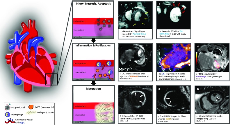
Summary: This review summarizes the state of art of non-invasive molecular imaging of the myocardium during ischemia and after myocardial infarction using PET and MRI. We also describe the different contrast agents that have been developed to image the different phases of cardiac healing and the biological processes associated with each of those phases. Importantly, here we focus on imaging of inflammation as it is the key biological process that orchestrates clearance of dead cells, tissue remodeling, cardiac repair, and future outcome. We also focus on clinical translation of some of the novel contrast agents that have been tested in patients and discuss the need for larger, multi-center patient studies to fully validate the applicability of new imaging probes.
Keywords: Cardiovascular imaging; Inflammation; Ischemic heart disease; Myocardial infarction; Vascular remodeling.
Conflict of interest statement
Conflict of InterestAll authors declare that they have no conflict of interest.
Figures


References
-
- Writing Group Members et al. Heart disease and stroke statistics-2016 update: a report from the American Heart Association. Circulation. 2016;133(4):e38–e60. - PubMed
-
- van der Laan AM, Nahrendorf M, Piek JJ. Healing and adverse remodelling after acute myocardial infarction: role of the cellular immune response. Heart. 2012;98(18):1384–1390. - PubMed
-
- McMurray JJ, Pfeffer MA. Heart failure. Lancet. 2005;365(9474):1877–1889. - PubMed
Publication types
Grants and funding
LinkOut - more resources
Full Text Sources
Research Materials
Miscellaneous