Review
. 2019 May;9(5):842-857.
doi: 10.21037/qims.2019.05.06.

Imaging the aged brain: pertinence and methods

Affiliations
Review

Imaging the aged brain: pertinence and methods

Hannah Humayun et al. Quant Imaging Med Surg. 2019 May.

Abstract

The global population is ageing at an accelerating speed. The ability to perform working memory tasks together with rapid processing becomes increasingly difficult with increases in age. With increasing national average life spans and a rise in the prevalence of age-related disease, it is pertinent to discuss the unique perspectives that can be gained from imaging the aged brain. Differences in structure, function, blood flow, and neurovascular coupling are present in both healthy aged brains and in diseased brains and have not yet been explored to their full depth in contemporary imaging studies. Imaging methods ranging from optical imaging to magnetic resonance imaging (MRI) to newer technologies such as photoacoustic tomography each offer unique advantages and challenges in imaging the aged brain. This paper will summarize first the importance and challenges of imaging the aged brain and then offer analysis of potential imaging modalities and their representative applications. The potential breakthroughs in brain imaging are also envisioned.

Keywords: Brain imaging; aging; magnetic resonance imaging (MRI); neuroscience; neurovascular coupling; optical imaging; positron emission tomography.


Conflict of interest statement

Conflicts of Interest: The authors have no conflicts of interest to declare.

Figures

Figure 1
Figure 1
Horizontal slice magnetic resonance imaging scans of a healthy 20-year-old brain (left) and a healthy 86-year-old brain (right). Side by side comparison to show the anatomical changes to brain structure in only the presence of aging. Notable differences occur in the widening of the third ventricle, the left lateral ventricle, the vermian subarachnoid space, and left circular sulcus. Widening can be noted by the greater presence of dark space on the scans.
Figure 2
Figure 2
Extrapolated points of difference between younger brains and older brains during functional neuroimaging study completed by Reuter-Lorenz and Lustig (62). Regions of impairment indicate loss of function with age and areas of compensation (usually paired with regions of impairment) are areas of higher activation in older brains trying to operate at a functional level in the task.
Figure 3
Figure 3
Graphical representation of memory functions that either stay stable or decline with age.
Figure 4
Figure 4
Illustration of the brain hierarchy, showing (A) the functional units at different scales and (B) the connections at different brain regions.
Figure 5
Figure 5
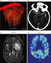
Representative brain imaging technologies. (A) Two-photon microscopy of dendrites of pyramidal neurons (shown in yellow) and blood vessels (shown in red) in somatosensory cortex of the Thy1-mitoCFP and Thy1-YFP mice. Adapted with permissions from (108). (B) Timing-invariant CT angiography image in a patient with a left-sided middle cerebral artery occlusion in the M2 segment. Adapted with permissions from (109). (C) Brain MRI: axial T2-weighted image of the brain demonstrates a hyperintense infiltrating mass of the left posterior frontal and temporal lobe with mass effect. Normal flow void is also noted through the left MCA branches. Adapted with permissions from (110). (D) PET scan in the parietal region in one subject with mild cognitive impairment who was reclassified on follow-up as having Alzheimer’s disease. Red and yellow areas correspond to high amyloid senile plaque values. Adapted with permissions from (111).

References

    1. Ezeh AC, Bongaarts J, Mberu B. Global population trends and policy options. Lancet 2012;380:142-8. 10.1016/S0140-6736(12)60696-5 - DOI - PubMed
    1. Wasay M, Grisold W, Carroll W, Shakir R. World Brain Day 2016: celebrating brain health in an ageing population. Lancet Neurol 2016;15:1008. 10.1016/S1474-4422(16)30171-5 - DOI - PubMed
    1. Wen YM. Health in an Aging World: What Should We Do? Engineering 2016;2:40-3. 10.1016/J.ENG.2016.01.010 - DOI
    1. Suzman R, Beard JR, Boerma T, Chatterji S. Health in an ageing world-what do we know? Lancet 2015;385:484. 10.1016/S0140-6736(14)61597-X - DOI - PubMed
    1. Ferri CP, Schoenborn C, Kalra L, Acosta D, Guerra M, Huang YQ, Jacob KS, Rodriguez JJL, Salas A, Sosa AL, Williams JD, Liu ZR, Moriyama T, Valhuerdi A, Prince MJ. Prevalence of stroke and related burden among older people living in Latin America, India and China. J Neurol Neurosurg Psychiatry 2011;82:1074-82. 10.1136/jnnp.2010.234153 - DOI - PMC - PubMed