Review
. 2019 May;9(5):905-920.
doi: 10.21037/qims.2019.05.16.

Advanced endoscopic methods in gastrointestinal diseases: a systematic review

Affiliations
Review

Advanced endoscopic methods in gastrointestinal diseases: a systematic review

Hua Li et al. Quant Imaging Med Surg. 2019 May.

Abstract

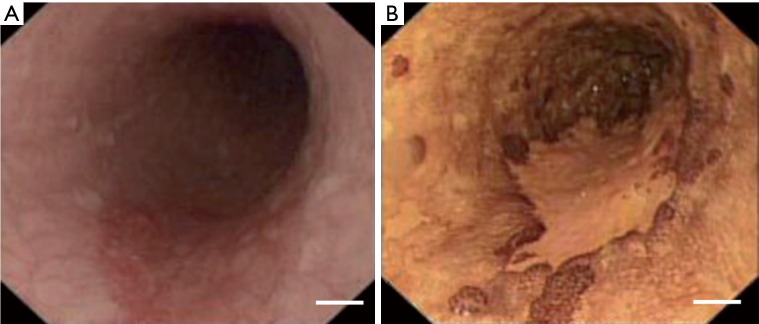
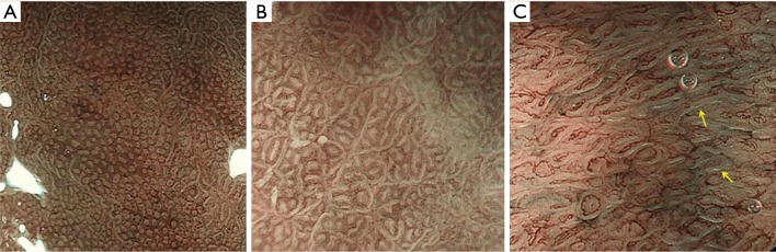
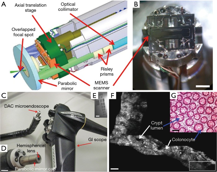
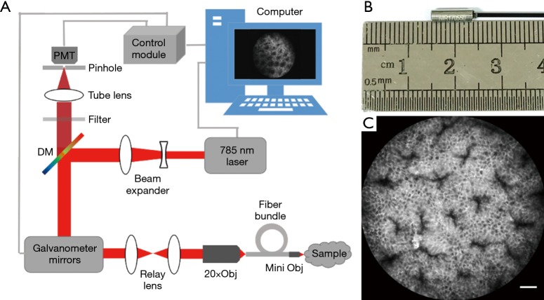
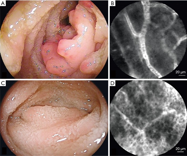
Endoscopic imaging is the main method for detecting gastrointestinal diseases, which adversely affect human health. White light endoscopy (WLE) was the first method used for endoscopic examination and is still the preliminary step in the detection of gastrointestinal diseases during clinical examination. However, it cannot accurately diagnose gastrointestinal diseases owing to its poor correlation with histopathological diagnosis. In recent years, many advanced endoscopic methods have emerged to improve the detection accuracy by endoscopy. Chromoendoscopy (CE) enhances the contrast between normal and diseased tissues using biocompatible dye agents. Narrow band imaging (NBI) can improve the contrast between capillaries and submucosal vessels by changing the light source acting on the tissue using special filters to realize the visualization of the vascular structure. Flexible spectral imaging color enhancement (FICE) technique uses the reflectance spectrum estimation technique to obtain individual spectral images and reconstructs an enhanced image of the mucosal surface using three selected spectral images. The i-Scan technology takes advantage of the different reflective properties of normal and diseased tissues to obtain images, and enhances image contrast through post-processing algorithms. These abovementioned methods can be used to detect gastrointestinal diseases by observing the macroscopic structure of the digestive tract mucosa, but the ability of early cancer detection is limited with low resolution. However, based on the principle of confocal imaging, probe-based confocal laser endomicroscopy (pCLE) can enable cellular visualization with high-performance probes, which can present cellular morphology that is highly consistent with that shown by biopsy to provide the possibility of early detection of cancer. Other endoscopic imaging techniques including endoscopic optical coherence tomography (EOCT) and photoacoustic endoscopy (PAE), are also promising for diagnosing gastrointestinal diseases. This review focuses on these technologies and aims to provide an overview of different technologies and their clinical applicability.

Keywords: Endoscopy; narrow band imaging (NBI); probe-based confocal laser endomicroscopy (pCLE).


Conflict of interest statement

Conflicts of Interest: The authors have no conflicts of interest to declare.

Figures

Figure 1
Figure 1
Images of the esophagus. (A) White light endoscopy; (B) chromoendoscopy using Lugol’s solution for a patch of high-grade dysplastic squamous epithelium. The dysplastic area remained unstained, whereas normal squamous epithelium shows coloration. Scale bar =2 cm. Reproduced with permission from (12).
Figure 2
Figure 2
Images of magnifying endoscopy (ME) with narrow-band imaging (NBI). (A) An abnormal form and arrangement of the crypt openings is observed in the fundic gland mucosa with inflammation; (B) elliptical or groove-like shapes with white color is observed in the fundic gland mucosa with inflammation; (C) atrophic mucosa of the corpus. Yellow arrows indicate a light blue crest (24).
Figure 3
Figure 3
Dual-axis confocal laser endomicroscope. (A) Two collimated beams are focused by a parabolic mirror; (B) photograph of a 2D microelectromechanical system (MEMS) scanner mounted on the axial translation stage; scale bar =3 mm; (C) a dual-axis confocal laser endomicroscope is passed through the instrument channel of an endoscope; (D) the distal end of dual-axis confocal laser endomicroscope; scale bar =5 mm; (E) lateral resolution is 5 µm; scale bar =20 µm; (F) a mosaic large field of view en face image of normal colonic mucosa at a depth of 60 µm and (G) a histologic (H&E) image of normal colonic mucosa. Scale bar =100 µm. The white rectangle represents an individual en face image (362 µm × 134 µm) obtained using a dual-axis confocal laser endomicroscope (47).
Figure 4
Figure 4
Homemade 785-nm near-infrared probe-based confocal laser endomicroscope. (A) Schematic diagram of probe-based confocal laser endomicroscope; (B) probe of the endomicroscope; (C) imaging of normal colon mucosa of C57BL/6 mice after intravenous ICG (15 mg/kg). Scale bar =30 µm.
Figure 5
Figure 5
Follicular lymphoma in the ileum. (A) Enteroscopic view before treatment; (B) “Soccer ball-like pattern” capillary vessels can be observed in a probe-based confocal laser endomicroscopic image; (C) enteroscopic view after rituximab monotherapy; (D) a probe-based confocal laser endomicroscopic image doesn’t show a “soccer ball-like pattern” capillary vessels after rituximab monotherapy (73).

References

    1. Siegel RL, Miller KD, Jemal A. Cancer statistics, 2018. CA Cancer J Clin 2018;68:7-30. 10.3322/caac.21442 - DOI - PubMed
    1. Atkins L, Benedict EB. Correlation of gross gastroscopic findings with gastroscopic biopsy in gastritis. N Engl J Med 1956;254:641-4. 10.1056/NEJM195604052541403 - DOI - PubMed
    1. Kato T, Yagi N, Kamada T, Shimbo T, Watanabe H, Ida K. Diagnosis of Helicobacter pylori infection in gastric mucosa by endoscopic features: A multicenter prospective study. Dig Endosc 2013;25:508-18. 10.1111/den.12031 - DOI - PubMed
    1. Cho JH, Chang YW, Jang JY, Shim JJ, Lee CK, Dong SH, Kim HJ, Kim B-H, Lee TH, Cho JY. Close observation of gastric mucosal pattern by standard endoscopy can predict Helicobacter pylori infection status. J Gastroenterol Hepatol 2013;28:279-84. 10.1111/jgh.12046 - DOI - PubMed
    1. Panteris V, Nikolopoulou S, Lountou A, Triantafillidis JK. Diagnostic capabilities of high-definition white light endoscopy for the diagnosis of gastric intestinal metaplasia and correlation with histologic and clinical data. Eur J Gastroenterol Hepatol 2014;26:594-601. - PubMed