. 2019 Jun 9:2019:5683479.
doi: 10.1155/2019/5683479. eCollection 2019.

Ischemic Postconditioning (IPostC) Protects Fibrotic and Cirrhotic Rat Livers after Warm Ischemia

Affiliations

Ischemic Postconditioning (IPostC) Protects Fibrotic and Cirrhotic Rat Livers after Warm Ischemia

Julia Schewe et al. Can J Gastroenterol Hepatol. .

Abstract

Background: Decreased organ function following liver resection is a major clinical issue. The practical method of ischemic postconditioning (IPostC) has been studied in heart diseases, but no data exist regarding fibrotic livers.

Aims: We aimed to determine whether IPostC could protect healthy, fibrotic, and cirrhotic livers from ischemia reperfusion injury (IRI).

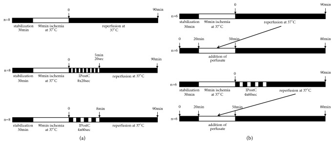
Methods: Fibrosis was induced in male SD rats using bile duct ligation (BDL, 4 weeks), and cirrhosis was induced using thioacetamide (TAA, 18 weeks). Fibrosis and cirrhosis were histologically confirmed using HE and EvG staining. For healthy, fibrotic, and cirrhotic livers, isolated liver perfusion with 90 min of warm ischemia was performed in three groups (each with n=8): control, IPostC 8x20 sec, and IPostC 4x60 sec. additionally, healthy livers were investigated during a follow-up study. Lactate dehydrogenase (LDH) and thromboxane B2 (TXB2) in the perfusate, as well as bile flow (healthy/TAA) and portal perfusion pressure, were measured.

Results: LDH and TXB2 were reduced, and bile flow was increased by IPostC, mainly in total and in the late phase of reperfusion. The follow-up study showed that the perfusate derived from a postconditioned group had much less damaging potential than perfusate derived from the nonpostconditioned group.

Conclusion: IPostC following warm ischemia protects healthy, fibrotic, and cirrhotic livers against IRI. Reduced efflux of TXB2 is one possible mechanism for this effect of IPostC and increases sinusoidal microcirculation. These findings may help to improve organ function and recovery of patients after liver resection.


Figures

Figure 1
Figure 1
Experimental protocols for animal studies. (a) Male SD rats underwent bile duct ligation over 4 weeks to induce liver fibrosis or received thioacetamide via drinking water over 18 weeks to induce liver cirrhosis. For the implementation of warm ischemia, healthy, fibrotic, and cirrhotic livers were perfused for 30 min, stored for 90 min at 37°C, and reperfused for 90 min. The IPostC intervention groups included 8x20 sec and 4x60 sec cycles that were performed after ischemia (control and intervention groups, each n=8). (b) For the follow-up study, we performed warm ischemia in healthy livers of male SD rats (postconditioned with IPostC 4x60 sec and nonpostconditioned, each n=6) and collected the reperfusion perfusate to perfuse a second healthy liver for 80 min.
Figure 2
Figure 2
Histological evaluation of healthy, fibrotic, and cirrhotic livers after warm ischemia. After the induction of liver fibrosis in male SD rats using bile duct ligation (BDL) and the induction of liver cirrhosis using thioacetamide (TAA) administered via drinking water, healthy (a/b), fibrotic (c/d), and cirrhotic (e/f) livers were perfused (100x magnification). A stabilization period of 30 min was followed by 90 min of storage at 37°C and 90 min of reperfusion. For the IPostC intervention groups, 8x20 sec and 4x60 sec cycles were performed after ischemia (control and intervention groups, each with n=8). After the experiments, liver samples were fixed in 4% formalin and histologically evaluated (Table 1).
Figure 3
Figure 3
Basal values of lactate dehydrogenase, bile flow, and portal perfusion pressure in healthy, fibrotic, and cirrhotic livers. To induce liver fibrosis, bile duct ligation (BDL) was performed in male SD rats, and to induce liver cirrhosis, animals received thioacetamide (TAA) administered via drinking water. Healthy, fibrotic, and cirrhotic livers were perfused for 30 min. Lactate dehydrogenase (LDH; (a)) was measured in the perfusate, bile was collected to determine the bile flow (b), and the portal perfusion pressure (c) was monitored continuously. All data are expressed as the mean ± SEM. Significant values are specified as ∗∗p<0.01 (fibrotic/cirrhotic liver compared to healthy liver) and ##p<0.01 (cirrhotic liver compared with fibrotic liver).
Figure 4
Figure 4
Lactate dehydrogenase, bile flow, and portal perfusion pressure in healthy, fibrotic, and cirrhotic livers after warm ischemia ± IPostC. Liver fibrosis was induced in male SD rats using bile duct ligation (BDL) over 4 weeks. Liver cirrhosis was induced using thioacetamide (TAA) administered via drinking water over 18 weeks. The healthy, fibrotic, and cirrhotic livers were perfused for 30 min, followed by 90 min of ischemia at 37°C and 90 min of reperfusion. The IPostC intervention groups included 8x20 sec and 4x60 sec cycles that were performed after ischemia (control and intervention groups, each n=8). Lactate dehydrogenase (LDH) was measured in the perfusate and is represented as total value (a) and during the early phase (minute 0 to 40; (b)) and the late phase (minute 50 to 90; (c)) of reperfusion. Bile was collected and also the bile flow is shown as total value (d) and at the early (e) and late (f) phase of reperfusion. For the portal perfusion pressure, which was monitored continuously, the maximum (g) and the value at the end of perfusion (h) are displayed. All data are expressed as the mean ± SEM, and significant values are specified as p<0.05 and ∗∗p<0.01.
Figure 5
Figure 5
Lactate dehydrogenase, bile flow, and portal perfusion pressure in healthy livers after follow-up study ± IPostC. Warm ischemia in situ ± IPostC 4x60 sec was performed in the healthy livers of male SD rats. The perfusate of reperfusion was collected and subsequently used to perfuse additional healthy livers (postconditioned and nonpostconditioned, each with n=6). Lactate dehydrogenase (LDH) was measured in the perfusate and bile was collected during the time of perfusion. Both LDH and bile flow are shown as maximal (LDH (a); bile (c)) and total (LDH (b); bile (d)) values. The portal perfusion pressure (e) was monitored continuously and is represented as maximal value. Data are expressed as the mean ± SEM. Significant values are specified as p<0.05 and ∗∗p<0.01.
Figure 6
Figure 6
Thromboxane B 2 in healthy, fibrotic, and cirrhotic livers after warm ischemia ± IPostC. The induction of liver fibrosis using bile duct ligation (BDL) over 4 weeks or the induction of liver cirrhosis using thioacetamide (TAA) administered via drinking water over 18 weeks in male SD rats was performed. The healthy, fibrotic, and cirrhotic livers were perfused for 30 min and stored at 37°C for 90 min, followed by reperfusion for 90 min. After ischemia, IPostC interventions using 8x20 sec and 4x60 sec (control and intervention groups, each with n=8) were carried out. Thromboxane B2 (TXB2) was measured in the collected perfusate using an enzyme-linked immunosorbent assay. The values for TXB2 are shown as basal values (a), in total (b) and during the early phase (minute 0 to 40; (c)) and the late phase (minute 50 to 90; (d)) of reperfusion. All data are expressed as the mean ± SEM. Significant values are specified as p<0.05 and ∗∗p<0.01.

References

    1. Mendes-Braz M., Elias-Miró M., Jiménez-Castro M. B., Casillas-Ramírez A., Ramalho F. S., Peralta C. The current state of knowledge of Hepatic Ischemia-ReperfusionInjury Based on Its Study in Experimental Models. Journal of Biomedicine and Biotechnology. 2012;2012:20. doi: 10.1155/2012/298657.298657 - DOI - PMC - PubMed
    1. Abu-Amara M., Gurusamy K. S., Hori S., Glantzounis G., Fuller B., Davidson B. R. Pharmacological interventions for ischaemia reperfusion injury in liver resection surgery performed under vascular control. Cochrane Database of Systematic Reviews. 2009;4 doi: 10.1002/14651858.CD008154.CD007472 - DOI - PMC - PubMed
    1. Saidi R. F., Chang J., Verb S., et al. The effect of methylprednisolone on warm ischemia-reperfusion injury in the liver. The American Journal of Surgery. 2007;193(3):345–348. doi: 10.1016/j.amjsurg.2006.09.017. - DOI - PubMed
    1. Soltys K., Dikdan G., Koneru B. Oxidative stress in fatty livers of obese Zucker rats: Rapid amelioration and improved tolerance to warm ischemia with tocopherol. Hepatology. 2001;34(1):13–18. doi: 10.1053/jhep.2001.25452. - DOI - PubMed
    1. Murry C. E., Jennings R. B., Reimer K. A. Preconditioning with ischemia: a delay of lethal cell injury in ischemic myocardium. Circulation. 1986;74(5):1124–1136. doi: 10.1161/01.CIR.74.5.1124. - DOI - PubMed