Comparative Study
. 2019 Jun 12:2019:2547216.
doi: 10.1155/2019/2547216. eCollection 2019.

Early Detection of Microvascular Changes in Patients with Diabetes Mellitus without and with Diabetic Retinopathy: Comparison between Different Swept-Source OCT-A Instruments

Affiliations
Comparative Study

Early Detection of Microvascular Changes in Patients with Diabetes Mellitus without and with Diabetic Retinopathy: Comparison between Different Swept-Source OCT-A Instruments

Stela Vujosevic et al. J Diabetes Res. .

Abstract

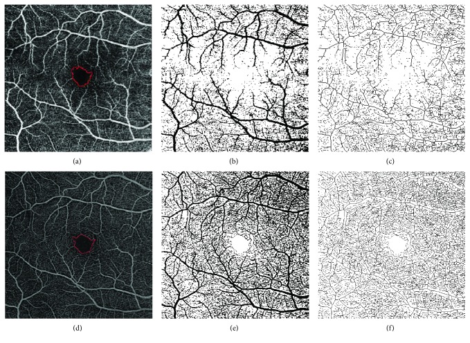
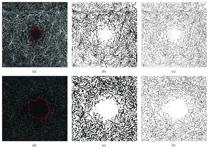
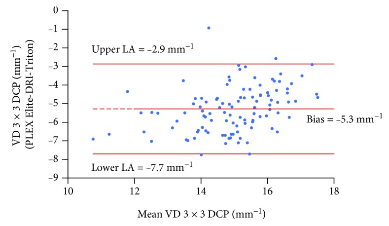
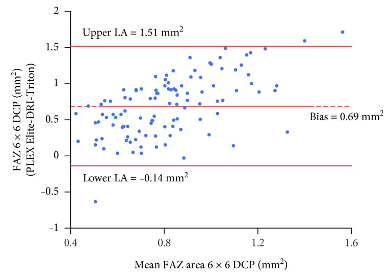
Optical coherence tomography angiography (OCT-A) has recently improved the ability to detect subclinical and early clinically visible microvascular changes occurring in patients with diabetes mellitus (DM). The aim of the present study is to evaluate and compare early quantitative changes of macular perfusion parameters in patients with DM without DR and with mild nonproliferative DR (NPDR) evaluated by two different swept-source (SS) OCT-A instruments using two scan protocols (3 × 3 mm and 6 × 6 mm). One hundred eleven subjects/eyes were prospectively evaluated: 18 healthy controls (control group), 73 eyes with DM but no DR (no-DR group), and 20 eyes with mild NPDR (DR group). All quantitative analyses were performed using ImageJ and included vessel and perfusion density, area and circularity index of the FAZ, and vascular complexity parameters. The agreement between methods was assessed according to the method of Bland-Altman. A significant decrease in the majority of the considered parameters was found in the DR group versus the controls with both instruments. The results of Bland-Altman analysis showed the presence of a systemic bias between the two instruments with PLEX Elite providing higher values for the majority of the tested parameters when considering 6 × 6 mm angiocubes and a less definite difference in 3 × 3 mm angiocubes. In conclusion, this study documents early microvascular changes occurring in the macular region of patients at initial stages of DR, confirmed with both SS OCT-A instruments. The fact that early microvascular alterations could not be detected with one instrument does not necessarily mean that these alterations are not actually present, but this could be an intrinsic limitation of the device itself. Further, larger longitudinal studies are needed to better understand microvascular damage at very early stages of diabetic retinal disease and to define the strengths and weaknesses of different OCT-A devices.


Figures

Figure 1
Figure 1
ImageJ analysis of 6 × 6 mm images at the SCP of a patient with DM and without DR. (a–c) SCP image obtained with DRI OCT-A Triton Plus (Topcon Medical Systems Europe, Milano, Italy). (d–f) SCP image obtained with prototype PLEX Elite 9000 (Carl Zeiss Meditec, Inc., Dublin, California, USA). (a, d) Original SCP slabs in which the FAZ profile was manually outlined using the freehand selections tool. (b, e) Binarized images. (c, f) Skeletonized images. SCP: superficial capillary plexus; DM: diabetes mellitus; DR: diabetic retinopathy; OCT-A: OCT angiography; FAZ: foveal avascular zone.
Figure 2
Figure 2
ImageJ analysis of 3 × 3 mm images at the DCP of a patient with DR. (a–c) DCP image obtained with DRI OCT-A Triton Plus (Topcon Medical Systems Europe, Milano, Italy). (d–f) DCP image obtained with prototype PLEX Elite 9000 (Carl Zeiss Meditec, Inc., Dublin, California, USA). (a, d) Original DCP slabs in which the FAZ profile was manually outlined using the freehand selections tool. (b, e) Binarized images. (c, f) Skeletonized images. DCP: deep capillary plexus; DR: diabetic retinopathy; OCT-A: OCT angiography; FAZ: foveal avascular zone.
Figure 3
Figure 3
Bland-Altman plot for VD in 3 × 3 mm angiocube scans evaluated at the DCP measured with PLEX Elite and DRI-Triton. The central line indicates the mean of the differences or bias; the upper and lower lines indicate the upper and lower limits of agreement (LA), respectively. VD: vessel density; DCP: deep capillary plexus.
Figure 4
Figure 4
Bland-Altman plot for the FAZ area in 6 × 6 mm angiocube scans evaluated at the DCP measured with PLEX Elite and DRI-Triton. The central line indicates the mean of the differences or bias; the upper and lower lines indicate the upper and lower limits of agreement (LA), respectively. DCP: deep capillary plexus.

References

    1. Yau J. W. Y., Rogers S. L., Kawasaki R., et al. Global prevalence and major risk factors of diabetic retinopathy. Diabetes Care. 2012;35(3):556–564. doi: 10.2337/dc11-1909. - DOI - PMC - PubMed
    1. Gardner T. W., Abcouwer S. F., Barber A. J., Jackson G. R. An integrated approach to diabetic retinopathy research. Archives of Ophthalmology. 2011;129(2):230–235. doi: 10.1001/archophthalmol.2010.362. - DOI - PMC - PubMed
    1. Joussen A. M., Poulaki V., Qin W., et al. Retinal vascular endothelial growth factor induces intercellular adhesion molecule-1 and endothelial nitric oxide synthase expression and initiates early diabetic retinal leukocyte adhesion in vivo. The American Journal of Pathology. 2002;160(2):501–509. doi: 10.1016/S0002-9440(10)64869-9. - DOI - PMC - PubMed
    1. Martin P. M., Roon P., van Ells T. K., Ganapathy V., Smith S. B. Death of retinal neurons in streptozotocin-induced diabetic mice. Investigative Opthalmology & Visual Science. 2004;45(9):3330–3336. doi: 10.1167/iovs.04-0247. - DOI - PubMed
    1. Lecleire-Collet A., Tessier L. H., Massin P., et al. Advanced glycation end products can induce glial reaction and neuronal degeneration in retinal explants. British Journal of Ophthalmology. 2005;89(12):1631–1633. doi: 10.1136/bjo.2005.079491. - DOI - PMC - PubMed

Publication types