Review
. 2020 Mar 20;30(4):226-240.
doi: 10.1093/glycob/cwz048.

Monitoring of immunoglobulin N- and O-glycosylation in health and disease

Affiliations
Review

Monitoring of immunoglobulin N- and O-glycosylation in health and disease

Noortje de Haan et al. Glycobiology. .

Abstract

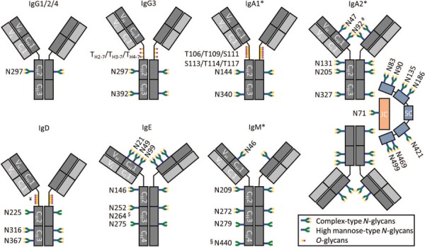
Protein N- and O-glycosylation are well known co- and post-translational modifications of immunoglobulins. Antibody glycosylation on the Fab and Fc portion is known to influence antigen binding and effector functions, respectively. To study associations between antibody glycosylation profiles and (patho) physiological states as well as antibody functionality, advanced technologies and methods are required. In-depth structural characterization of antibody glycosylation usually relies on the separation and tandem mass spectrometric (MS) analysis of released glycans. Protein- and site-specific information, on the other hand, may be obtained by the MS analysis of glycopeptides. With the development of high-resolution mass spectrometers, antibody glycosylation analysis at the intact or middle-up level has gained more interest, providing an integrated view of different post-translational modifications (including glycosylation). Alongside the in-depth methods, there is also great interest in robust, high-throughput techniques for routine glycosylation profiling in biopharma and clinical laboratories. With an emphasis on IgG Fc glycosylation, several highly robust separation-based techniques are employed for this purpose. In this review, we describe recent advances in MS methods, separation techniques and orthogonal approaches for the characterization of immunoglobulin glycosylation in different settings. We put emphasis on the current status and expected developments of antibody glycosylation analysis in biomedical, biopharmaceutical and clinical research.

Keywords: antibody; biopharmaceutical; glycan; glycoproteomics; mass spectrometry.


Figures

Fig. 1
Fig. 1
Schematic representation of the glycosylation sites of IgG, IgA, IgD, IgE and IgM. The IgG glycosylation sites are indicated by their amino acid number according to literature, e.g. Arnold et al. 2007 and Plomp et al. 2015, while the other isotypes follow UniProt numbering (for an overview of Ig glycosylation site nomenclature, see Plomp et al. 2016). Each Ig monomer is composed of two heavy chains (dark gray) and two light chains (light gray), connected by disulfide bonds. The chains are further subdivided as constant (C) and variable (V) domains. Polyclonal immunoglobulins may carry additional occupied glycosylation sites in their (hyper) variable regions in the Fab domain, which are not indicated in this figure. *IgA2 is shown in its dimeric form and in complex with the joining chain (JC; red) and the secretory component (SC; blue; present only for secretory IgA), both linked via disulfide bonds. Although not shown here, also IgA1 can form dimers, while IgM generally forms pentamers, both in combination with one joining chain. #N92 on IgA2 is only incorporated in an N-glycosylation consensus sequence in the IgAn and IgA2m(2) allotypes; N92 in IgA2m(1) is not glycosylated (Plomp et al. 2018; Chandler et al. 2019). $Although N264 of IgE is part of an N-glycosylation consensus sequence (NHS), this site was reported to be non-glycosylated when studied at the glycopeptide level (Plomp et al. 2014; Wu et al. 2016). ¥There are nine potential O-glycosylation sites present in the hinge region of IgD (S109, S10, T113, S121, T126, T127, T131, T131 and T135) for which different occupancies have been reported (Takayasu et al. 1982; Mellis and Baenziger 1983). §IgM N440 was reported to have a 30–40% site occupancy when derived from human plasma (Chandler et al. 2019). The green–blue–yellow structures at the glycosylation sites represent the presence of mainly complex type N-glycans, while the green–blue structures represent the presence of mainly high-mannose type N-glycans. The yellow–pink structures indicate the presence of O-glycosylation.
Fig. 2
Fig. 2
IgG Fc glycosylation is dependent on the expression system. Shown are the relative abundances of (A) high mannose-type glycans, (B) hybrid-type glycans, (C) galactosylation, (D) fucosylation, (E) the presence of a bisecting GlcNAc, (F) Neu5Ac sialylation, (G) Neu5Gc sialylation and (H) α1,3 galactosylation of human IgG1 (Plomp et al. 2017; Simurina et al. 2018), murine IgG1 (de Haan et al. 2017), Trastuzumab produced in CHO cells (Stadlmann et al. 2008), human IgG1 expressed in free style HEK cells (Dekkers et al. 2016), Cetuximab expressed in SP2/0 cells and Daclizumab expressed in NS0 cells (Stadlmann et al. 2008). Notably, while in most systems the sialylation is α2,6-linked, in CHO cells exclusively α2,3-linked sialylation is present. Next to the expression system, also biological effects (like age and disease for human IgG glycosylation and mouse strain for murine glycosylation) and the fermentation process (for cellular expression systems) have an effect on Fc glycosylation. For example, also Neu5Gc sialylated species have been reported on IgG expressed in NS0 cells (Montesino et al. 2012).

Similar articles

Cited by

References

    1. Abrahams JL, Campbell MP, Packer NH. 2018. Building a PGC-LC-MS N-glycan retention library and elution mapping resource. Glycoconj J. 35:15–29. - PubMed
    1. Ackerman ME, Crispin M, Yu X, Baruah K, Boesch AW, Harvey DJ, Dugast AS, Heizen EL, Ercan A, Choi I et al. . 2013. Natural variation in Fc glycosylation of HIV-specific antibodies impacts antiviral activity. J Clin Invest. 123:2183–2192. - PMC - PubMed
    1. Arnaud J, Audfray A, Imberty A. 2013. Binding sugars: From natural lectins to synthetic receptors and engineered neolectins. Chem Soc Rev. 42:4798–4813. - PubMed
    1. Arnold JN, Wormald MR, Sim RB, Rudd PM, Dwek RA. 2007. The impact of glycosylation on the biological function and structure of human immunoglobulins. Annu Rev Immunol. 25:21–50. - PubMed
    1. Bobaly B, D'Atri V, Lauber M, Beck A, Guillarme D, Fekete S. 2018. Characterizing various monoclonal antibodies with milder reversed phase chromatography conditions. J Chromatogr B Analyt Technol Biomed Life Sci. 1096:1–10. - PubMed

Publication types