

Drosophila Myoblast Fusion: Invasion and Resistance for the Ultimate Union
- PMID: 31283358
- PMCID: PMC7448821
- DOI: 10.1146/annurev-genet-120116-024603
Drosophila Myoblast Fusion: Invasion and Resistance for the Ultimate Union
Abstract
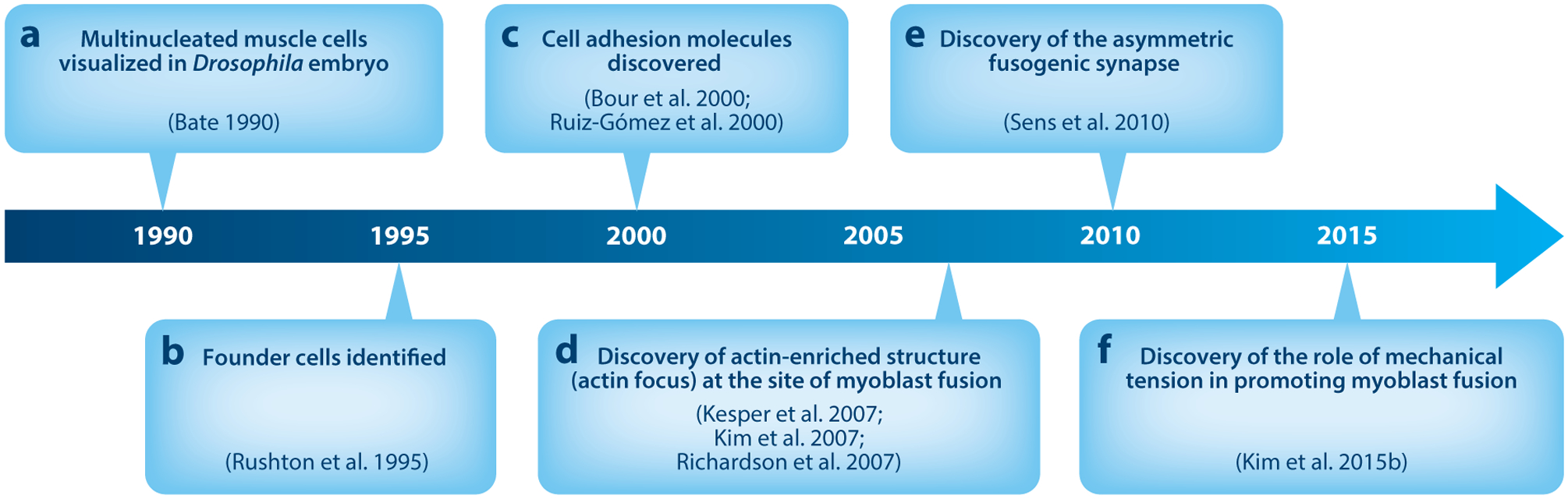
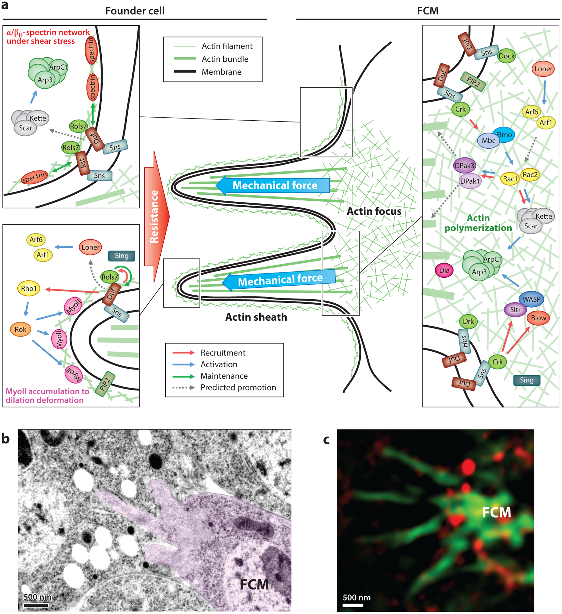
Cell-cell fusion is indispensable for creating life and building syncytial tissues and organs. Ever since the discovery of cell-cell fusion, how cells join together to form zygotes and multinucleated syncytia has remained a fundamental question in cell and developmental biology. In the past two decades, Drosophila myoblast fusion has been used as a powerful genetic model to unravel mechanisms underlying cell-cell fusion in vivo. Many evolutionarily conserved fusion-promoting factors have been identified and so has a surprising and conserved cellular mechanism. In this review, we revisit key findings in Drosophila myoblast fusion and highlight the critical roles of cellular invasion and resistance in driving cell membrane fusion.
Keywords: Drosophila genetics; actin-propelled membrane protrusions; asymmetric fusogenic synapse; cell–cell fusion; mechanical tension; mechanosensory response; myoblast fusion.
Figures
Similar articles
-
The fusogenic synapse at a glance.J Cell Sci. 2019 Sep 16;132(18):jcs213124. doi: 10.1242/jcs.213124. J Cell Sci. 2019. PMID: 31527149 Free PMC article. Review.
-
WIP/WASp-based actin-polymerization machinery is essential for myoblast fusion in Drosophila.Dev Cell. 2007 Apr;12(4):557-69. doi: 10.1016/j.devcel.2007.01.016. Dev Cell. 2007. PMID: 17419994
-
Asymmetric Mbc, active Rac1 and F-actin foci in the fusion-competent myoblasts during myoblast fusion in Drosophila.Development. 2011 Apr;138(8):1551-62. doi: 10.1242/dev.057653. Epub 2011 Mar 9. Development. 2011. PMID: 21389053 Free PMC article.
-
A conserved molecular pathway mediates myoblast fusion in insects and vertebrates.Nat Genet. 2007 Jun;39(6):781-6. doi: 10.1038/ng2055. Epub 2007 May 27. Nat Genet. 2007. PMID: 17529975
-
Drosophila melanogaster: A Model System to Study Distinct Genetic Programs in Myoblast Fusion.Cells. 2022 Jan 19;11(3):321. doi: 10.3390/cells11030321. Cells. 2022. PMID: 35159130 Free PMC article. Review.
Cited by
-
Cell Fusion: Merging Membranes and Making Muscle.Trends Cell Biol. 2019 Dec;29(12):964-973. doi: 10.1016/j.tcb.2019.09.002. Epub 2019 Oct 21. Trends Cell Biol. 2019. PMID: 31648852 Free PMC article. Review.
-
Cell Fusion-Related Proteins and Signaling Pathways, and Their Roles in the Development and Progression of Cancer.Front Cell Dev Biol. 2022 Feb 1;9:809668. doi: 10.3389/fcell.2021.809668. eCollection 2021. Front Cell Dev Biol. 2022. PMID: 35178400 Free PMC article. Review.
-
FGF signaling promotes spreading of fat body precursors necessary for adult adipogenesis in Drosophila.PLoS Biol. 2023 Mar 22;21(3):e3002050. doi: 10.1371/journal.pbio.3002050. eCollection 2023 Mar. PLoS Biol. 2023. PMID: 36947563 Free PMC article.
-
ERK1/2 inhibition promotes robust myotube growth via CaMKII activation resulting in myoblast-to-myotube fusion.Dev Cell. 2021 Dec 20;56(24):3349-3363.e6. doi: 10.1016/j.devcel.2021.11.022. Dev Cell. 2021. PMID: 34932950 Free PMC article.
-
Wound-Induced Syncytia Outpace Mononucleate Neighbors during Drosophila Wound Repair.bioRxiv [Preprint]. 2023 Oct 26:2023.06.25.546442. doi: 10.1101/2023.06.25.546442. bioRxiv. 2023. PMID: 37425719 Free PMC article. Preprint.
References
-
- Arias-Romero LE, Chernoff J. 2008. A tale of two Paks. Biol. Cell 100:97–108 - PubMed
-
- Artero R, Furlong EE, Beckett K, Scott MP, Baylies M. 2003. Notch and Ras signaling pathway effector genes expressed in fusion competent and founder cells during Drosophila myogenesis. Development 130:6257–72 - PubMed
-
- Artero RD, Castanon I, Baylies MK. 2001. The immunoglobulin-like protein Hibris functions as a dose-dependent regulator of myoblast fusion and is differentially controlled by Ras and Notch signaling. Development 128:4251–64 - PubMed
Publication types
MeSH terms
Substances
Grants and funding
LinkOut - more resources
Full Text Sources
Molecular Biology Databases
Miscellaneous
