Review
. 2019 Sep 29;6(1):501-524.
doi: 10.1146/annurev-virology-101416-041429. Epub 2019 Jul 5.

In Vivo Imaging-Driven Approaches to Study Virus Dissemination and Pathogenesis

Affiliations
Review

In Vivo Imaging-Driven Approaches to Study Virus Dissemination and Pathogenesis

Pradeep D Uchil et al. Annu Rev Virol. .

Abstract

Viruses are causative agents for many diseases and infect all living organisms on the planet. Development of effective therapies has relied on our ability to isolate and culture viruses in vitro, allowing mechanistic studies and strategic interventions. While this reductionist approach is necessary, testing the relevance of in vitro findings often takes a very long time. New developments in imaging technologies are transforming our experimental approach where viral pathogenesis can be studied in vivo at multiple spatial and temporal resolutions. Here, we outline a vision of a top-down approach using noninvasive whole-body imaging as a guide for in-depth characterization of key tissues, physiologically relevant cell types, and pathways of spread to elucidate mechanisms of virus spread and pathogenesis. Tool development toward imaging of infectious diseases is expected to transform clinical diagnosis and treatment.

Keywords: immune response; noninvasive imaging; virus dissemination; virus infection.


Figures
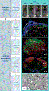
Figure 1
Figure 1
Illustration of a technological pipeline for a whole-body imaging-driven multiscale investigative approach to study virus infection. MLV is used as an example. (Top right) Longitudinal NBLI reveals infection of NLuc-expressing MLV at the pLN after subcutaneous challenge and informs subsequent focused studies at the pLN using various imaging modalities (middle column) at increasing scales of resolution (left column). (Right column) Intravital microscopy shows the arrival of fluorescent Gag-GFP-labeled MLV at the pLN (blue collagen capsule) and reveals trans-infectious synapses between MLV-laden SCS macrophages (green) and B-1 cells (red). Light sheet fluorescence microscopy shows the entire LN with MLV (green) captured at the pLN. Confocal immunohistochemistry of pLN sections shows MLV Gag-GFP (green) accumulating below collagen capsule (blue) next to B cell-rich follicles (red). Electron tomography reveals cell-cell contacts between B cells and MLV-laden SCS macrophages in LNs. Abbreviations: GFP, green fluorescent protein; LN, lymph node; MLV, murine leukemia virus; NBLI, noninvasive bioluminescence imaging; NLuc, Nanoluciferase; pLN, popliteal lymph node; SCS, subcapsular sinus. The images for intravital, confocal, and electron microscopy are reproduced from Reference with permission from AAAS.
Figure 2
Figure 2
General illustration showing spatiotemporal progression of virus infection in a mammalian host and the toolbox required to monitor virus-host interactions. (a) This illustration shows the various stages of virus infection from initial replication to systemic dissemination and its transit through various organs in the host after entering through indicated routes until its transmission from various shedding sites. (b) Shown are the indicated phases of virus infection and ensuing host immune responses that need to be monitored using the toolbox of viral as well as host-encoded reporters for in vivo imaging-guided studies of infection. Abbreviations: CMV, cytomegalovirus; GI, gastrointestinal; HCV, hepatitis C virus; HIV, human immunodeficiency virus; HPV, human papillomavirus; HSV, herpes simplex virus; IFN, interferon; JEV, Japanese encephalitis virus; WNV, West Nile virus; YFV, yellow fever virus.
Figure 3
Figure 3
Illustration showing the fate of blood- and lymph-borne viruses after capture by CD169-expressing MMMs in the splenic MZs and SCS-lining macrophages of the popliteal LN. MMMs and SCS macrophages play an infection-promoting role by capturing MLV particles from blood and lymph before trans-infecting permissive lymphocytes, such as B-1 cells. In the case of the pathogenic FVC, MMMs in the spleen limit virus dissemination to the red-pulp resident erythroblasts and orchestrate protective CD8+ T cell responses by collaborating with cDC1. MuHV-4 infects splenic MZ macrophages, followed by MZ B cells. The infected MZ B cells migrate to the white pulp, where they transfer virus to FDCs, which spread the infection to interacting B cells. In the LN, SCS macrophages restrict MuHV-4 spread by sequestering viruses from lymph. However, migratory DCs carry MuHV-4 from the upper respiratory tract to the LN and promote infection by spreading virus to B cells. MCMV infects SCS macrophages in the LN, which do not support robust virus infection, thus restricting viral dissemination to potential targets. Blood-borne VSV infects MMMs and red-pulp resident F4/80+ macrophages in the spleen. Unlike F4/80+ macrophages, MMMs are productively infected by VSV and serve as vessels for antigen and IFN production. The DC activation that follows helps in eliciting protective humoral and cell-mediated responses against VSV. Lymph-borne VSV infects SCS macrophages, which produce IFN to protect peripheral nerves from lethal VSV infection. Individual viruses are demarcated in the overview (left) using blue and white quadrants. Black arrows denote blood or lymph flow, and green arrows show the flow of viral particles. Virus-infected cells are shown in green, and red crosses depict restricted viral spread. Abbreviations: cDC1, conventional dendritic cells 1; CNS, central nervous system; DC, dendritic cell; FDC, follicular dendritic cell; FVC, Friend virus complex; GM3, monosialodihexosylganglioside; IFN, interferon; LN, lymph node; MCMV, murine cytomegalovirus; MLV, murine leukemia virus; MMM, marginal zone metallophilic macrophage; MuHV-4, murid herpesvirus 4; MZ, marginal zone; SCS, subcapsular sinus; VSV, vesicular stomatitis virus.

References

    1. Raoult D, Forterre P. 2008. Redefining viruses: lessons from Mimivirus. Nat. Rev. Microbiol 6:315–19 - PubMed
    1. Edwards RA, Rohwer F. 2005. Viral metagenomics. Nat. Rev. Microbiol 3:504–10 - PubMed
    1. Iwasaki A, Pillai PS. 2014. Innate immunity to influenza virus infection. Nat. Rev. Immunol 14:315–28 - PMC - PubMed
    1. Paules C, Subbarao K. 2017. Influenza. Lancet 390:697–708 - PubMed
    1. Sharp PM, Hahn BH. 2011. Origins of HIV and the AIDS pandemic. Cold Spring Harb. Perspect. Med 1:a006841. - PMC - PubMed

Publication types