

Mucinous Neoplasms of the Ovary: Radiologic-Pathologic Correlation
- PMID: 31283462
- PMCID: PMC6677283
- DOI: 10.1148/rg.2019180221
Mucinous Neoplasms of the Ovary: Radiologic-Pathologic Correlation
Abstract
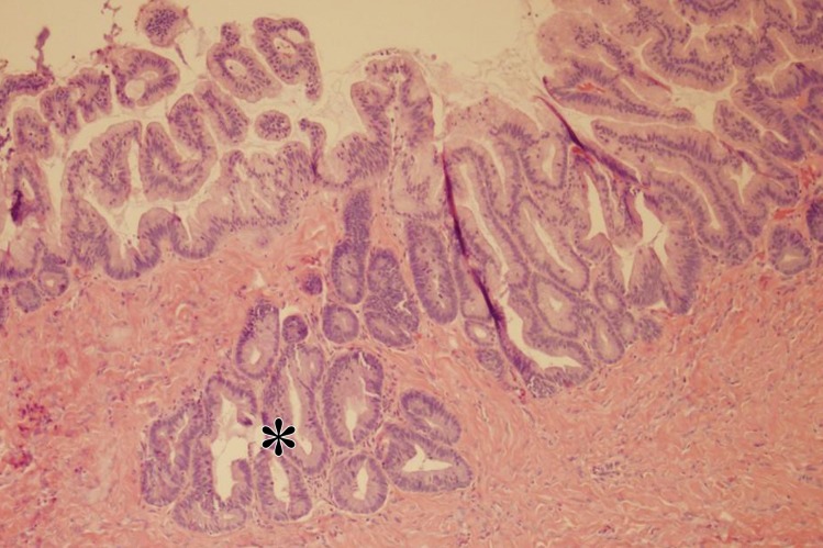
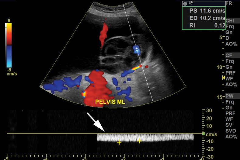
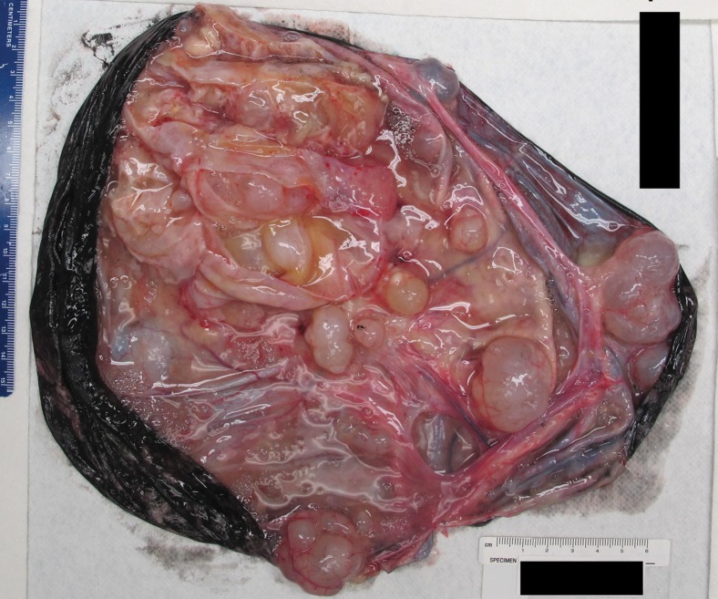
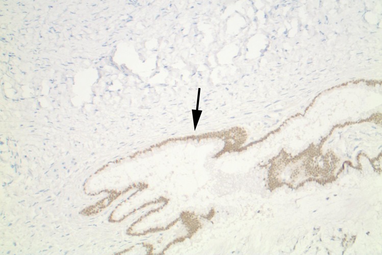
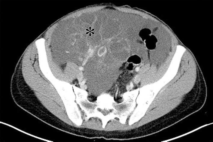
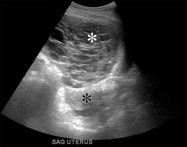
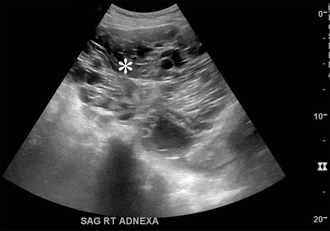
Mucinous neoplasms of the ovary account for 10%-15% of ovarian neoplasms. They may be benign, borderline, or malignant. The large majority are benign or borderline, accounting for 80% and 16%-17%, respectively. Mucinous neoplasms of the ovary most commonly affect women in their 20s to 40s. The clinical manifestation is nonspecific, but most mucinous ovarian neoplasms manifest as large unilateral pelvic masses. At gross pathologic analysis, mucinous ovarian neoplasms appear as large multiloculated cystic masses. The contents of the cyst loculi vary on the basis of differences in internal mucin content. At histologic analysis, mucinous ovarian neoplasms are composed of multiple cysts lined by mucinous epithelium, often resembling gastrointestinal-type epithelium. Imaging evaluation most commonly includes US and/or MRI. The imaging findings parallel the gross pathologic features and include a large, unilateral, multiloculated cystic mass. The cyst loculi vary in echogenicity, attenuation, and signal intensity depending on the mucin content. Mucinous neoplasms of the ovary are staged surgically using the FIGO (International Federation of Gynecology and Obstetrics) staging system. Primary treatment is surgical, with adjuvant chemotherapy considered in the uncommon case of mucinous carcinoma with extraovarian disease. Since most mucinous ovarian neoplasms are benign or borderline, the overall prognosis is excellent.
Figures


References
-
- Kurman RJ, Carcangiu ML, Herrington S, Young RH. World Health Organization classification of tumours of the female reproductive organs. 4th ed Lyon, France: IARC, 2014.
-
- Prat J, D’Angelo E, Espinosa I. Ovarian carcinomas: at least five different diseases with distinct histological features and molecular genetics. Hum Pathol 2018;80:11–27. - PubMed
-
- Lee KR, Young RH. The distinction between primary and metastatic mucinous carcinomas of the ovary: gross and histologic findings in 50 cases. Am J Surg Pathol 2003;27(3):281–292. - PubMed
-
- Berek JS, Hacker NF. Berek & Hacker’s gynecologic oncology. Philadelphia, Pa: Lippincott Williams & Wilkins, 2015.
-
- Riopel MA, Ronnett BM, Kurman RJ. Evaluation of diagnostic criteria and behavior of ovarian intestinal-type mucinous tumors: atypical proliferative (borderline) tumors and intraepithelial, microinvasive, invasive, and metastatic carcinomas. Am J Surg Pathol 1999;23(6):617–635. - PubMed
Publication types
MeSH terms
LinkOut - more resources
Full Text Sources
Medical
