

Light-Sheet Microscopy in Neuroscience
- PMID: 31283896
- PMCID: PMC6800245
- DOI: 10.1146/annurev-neuro-070918-050357
Light-Sheet Microscopy in Neuroscience
Abstract
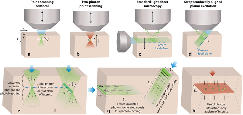
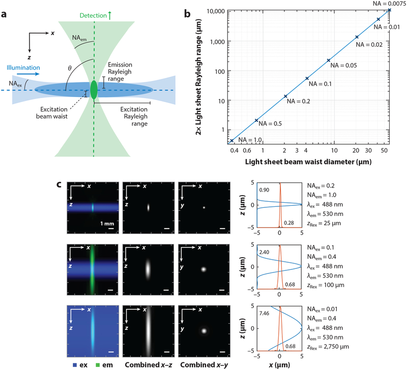
Light-sheet microscopy is an imaging approach that offers unique advantages for a diverse range of neuroscience applications. Unlike point-scanning techniques such as confocal and two-photon microscopy, light-sheet microscopes illuminate an entire plane of tissue, while imaging this plane onto a camera. Although early implementations of light sheet were optimized for longitudinal imaging of embryonic development in small specimens, emerging implementations are capable of capturing light-sheet images in freely moving, unconstrained specimens and even the intact in vivo mammalian brain. Meanwhile, the unique photobleaching and signal-to-noise benefits afforded by light-sheet microscopy's parallelized detection deliver the ability to perform volumetric imaging at much higher speeds than can be achieved using point scanning. This review describes the basic principles and evolution of light-sheet microscopy, followed by perspectives on emerging applications and opportunities for both imaging large, cleared, and expanded neural tissues and high-speed, functional imaging in vivo.
Keywords: GCaMP; functional imaging; light sheet; microscopy; tissue clearing.
Conflict of interest statement
DISCLOSURE STATEMENT
SCAPE intellectual property is licensed to Leica Microsystems for commercial development. All authors have a potential financial conflict of interest relating to SCAPE microscopy.
Figures
Similar articles
-
Light sheet fluorescence microscopy for neuroscience.J Neurosci Methods. 2019 May 1;319:16-27. doi: 10.1016/j.jneumeth.2018.07.011. Epub 2018 Jul 23. J Neurosci Methods. 2019. PMID: 30048674 Review.
-
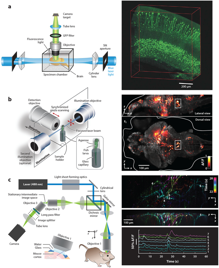
Swept confocally-aligned planar excitation (SCAPE) microscopy for high speed volumetric imaging of behaving organisms.Nat Photonics. 2015 Feb;9(2):113-119. doi: 10.1038/nphoton.2014.323. Nat Photonics. 2015. PMID: 25663846 Free PMC article.
-
Expansion Light Sheet Microscopy Resolves Subcellular Structures in Large Portions of the Songbird Brain.Front Neuroanat. 2019 Jan 31;13:2. doi: 10.3389/fnana.2019.00002. eCollection 2019. Front Neuroanat. 2019. PMID: 30766480 Free PMC article.
-
Light sheet microscopy of living or cleared specimens.Curr Opin Neurobiol. 2012 Feb;22(1):138-43. doi: 10.1016/j.conb.2011.08.003. Epub 2011 Sep 16. Curr Opin Neurobiol. 2012. PMID: 21925871 Review.
-
Volumetric Imaging of Neural Activity by Light Field Microscopy.Neurosci Bull. 2022 Dec;38(12):1559-1568. doi: 10.1007/s12264-022-00923-9. Epub 2022 Aug 8. Neurosci Bull. 2022. PMID: 35939199 Free PMC article. Review.
Cited by
-
Cryogenic contrast-enhanced microCT enables nondestructive 3D quantitative histopathology of soft biological tissues.Nat Commun. 2022 Oct 20;13(1):6207. doi: 10.1038/s41467-022-34048-4. Nat Commun. 2022. PMID: 36266273 Free PMC article.
-
A Model of Discovery: The Role of Imaging Established and Emerging Non-mammalian Models in Neuroscience.Front Mol Neurosci. 2022 Apr 14;15:867010. doi: 10.3389/fnmol.2022.867010. eCollection 2022. Front Mol Neurosci. 2022. PMID: 35493325 Free PMC article. Review.
-
Obesity-Related Neuroinflammation: Magnetic Resonance and Microscopy Imaging of the Brain.Int J Mol Sci. 2022 Aug 8;23(15):8790. doi: 10.3390/ijms23158790. Int J Mol Sci. 2022. PMID: 35955925 Free PMC article. Review.
-
Light-sheet autofluorescence lifetime imaging with a single-photon avalanche diode array.J Biomed Opt. 2023 Jun;28(6):066502. doi: 10.1117/1.JBO.28.6.066502. Epub 2023 Jun 21. J Biomed Opt. 2023. PMID: 37351197 Free PMC article.
-
Study liquid-liquid phase separation with optical microscopy: A methodology review.APL Bioeng. 2023 May 9;7(2):021502. doi: 10.1063/5.0137008. eCollection 2023 Jun. APL Bioeng. 2023. PMID: 37180732 Free PMC article. Review.
References
-
- Ahrens MB, Orger MB, Robson DN, Li JM, Keller PJ. 2013. Whole-brain functional imaging at cellular resolution using light-sheet microscopy. Nat. Methods 10:413–20 - PubMed
-
- Bewersdorf J, Pick R, Hell SW. 1998. Multifocal multiphoton microscopy. Opt. Lett 23:655–57 - PubMed
-
- Botcherby EJ, Juskaitis R, Booth MJ, Wilson T. 2007. Aberration-free optical refocusing in high numerical aperture microscopy. Opt. Lett 32:2007–9 - PubMed
Publication types
MeSH terms
Grants and funding
LinkOut - more resources
Full Text Sources
