. 2019 Jul 5;12(13):2157.
doi: 10.3390/ma12132157.

3D Human Periodontal Stem Cells and Endothelial Cells Promote Bone Development in Bovine Pericardium-Based Tissue Biomaterial

Affiliations

3D Human Periodontal Stem Cells and Endothelial Cells Promote Bone Development in Bovine Pericardium-Based Tissue Biomaterial

Jacopo Pizzicannella et al. Materials (Basel). .

Abstract

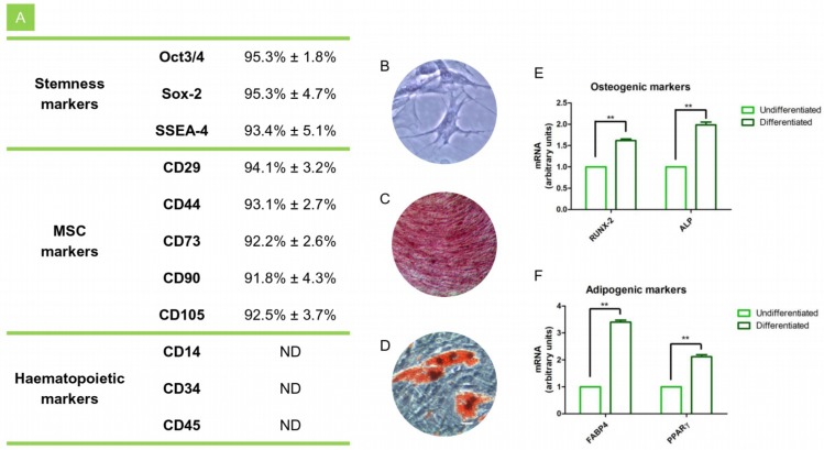
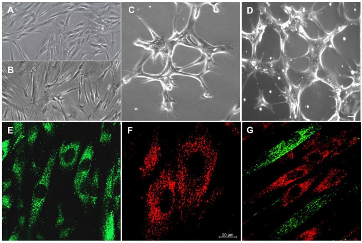
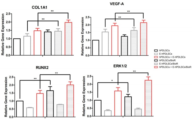
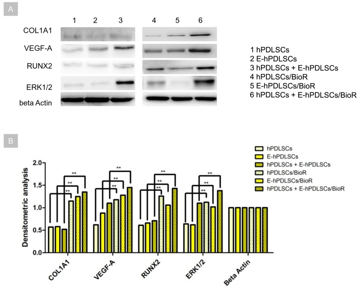
Bone defects repair represents a public and urgent problem in clinical practice, in fact, every year, more than two million patients required new treatments for bone injuries. Today a complete vascularization is strategic in bone formation, representing a new frontier for clinical application. Aim of this research has been developed a three-dimensional (3D) coculture platform using a bovine pericardium collagen membrane (BioR) loaded with human periodontal ligament stem cells (hPDLSCs) and endothelial differentiated cells from hPDLSCs (E-hPDLSCs) able to undergo toward osteoangiogenesis differentiation process. First, we have characterized at confocal laser scanning microscopy (CLSM) level the E-hPDLSCs phenotype profile, through CD31 and CD34 markers expression and the ability to tube vessel formation. Real Time-Polimerase Chain Reaction (RT-PCR) and western blotting analyses revealed the upregulation of Runt-related transcription factor 2 (RUNX2), Collagen 1A1 (COL1A1), Vascular Endothelial Growth Factor-A (VEGF-A) genes and proteins in the living construct composed by hPDLSCs + E-hPDSCs/BioR. Human PDLSCs + E-hPDLSCs/BioR construct showed also an enhacement of de novo synthesis of osteocalcin. Given that, the extracellular-signal-regulated kinase (ERK)/mitogen activated protein kinase (MAPK) transduction signaling was involved in the osteogenesis and angiogenesis process, the ERK1/2 protein level at biochemical level, in our experimental model, has been investigated. Our results evidenced an upregulation of ERK1/2 proteins level born in the living construct. In conclusion, we believe that the use of the hPDLSCs and E-hPDLSCs coculture togheter with BioR as substrate, could represent an efficient model able to activate through ERK1/2 signaling pathway the osteoangiogenesis process, and then representing a new potential engineered platform for surgeons during the repair and the healing of bone defects.

Keywords: Endothelial Cells differentiation; Runt-related transcription factor 2 (RUNX2); Vascular Endothelial Growth Factor-A (VEGF-A); bovine pericardium membrane; human Periodontal Ligament Stem Cells.


Conflict of interest statement

The authors declare no conflict of interest.

Figures

Figure 1
Figure 1
hPDLSCs characterization. (A) Cytofluorimetric analysis of hPDLSCs. (B) Plastic adherent hPDLSCs stained with toluidine blue solution. (C) Osteogenic differentiated hPDLSCs stained with Alizarin Red solution. (D) Adipogenic differentiated hPDLSCs stained with oil red solution. (E) RT-PCR of osteogenic related markers in undifferentiated and differentiated cells. (F) RT-PCR of adipogenic related markers in undifferentiated and differentiated cells. **p < 0.01. Scale bar: 10 µm.
Figure 2
Figure 2
Morphological analyses. (A) Undifferentiated hPDLSCs observed at light microscopy. (B) E-hPDLSCs observed at light microscopy. (C,D) E-hPDLSCs cultured on Cultrex® observed at light microscopy. (E) Undifferentiated hPDLSCs evaluated under CLSM (PKH67, green fluorescence). (F) E-hPDLSCs evaluated under at CLSM (PKH26, red fluorescence). (G) Coculture of hPDLSCs and E-hPDLSCs. Scale bar: 20 µm. Magnification: 10× (AD). Magnification: 20× (EG).
Figure 3
Figure 3
Immunofluorescence analyses. (A) Undifferentiated hPDLSCs showed a negative expression for CD31. (B) Undifferentiated hPDLSCs showed a negative expression for CD34. (C) Undifferentiated hPDLSCs showed a slightly positive expression for Vascular Endothelial Growth Factor-A (VEGF-A). (D) E-hPDLSCs showed a strong positive expression for CD31. (E) E-hPDLSCs showed a strong positive expression for CD34. (F) E-hPDLSCs showed a strong positive expression for VEGF-A. Scale bar: 10 µm. Magnification: 20×. Green fluorescent: cytoskeleton actin; Blue fluorescent: nuclei; Red fluorescent: specific markers. Cytofluorimetric analysis of the expression of CD31 and CD34 in hPDLSCs and in E-hPDLSCs confirm the immunofluorescence qualitative results.
Figure 4
Figure 4
Cell and 3D scaffold interaction. (A1) hPDLSCs, stained with PKH67 (green fluorescence), cultured on BioR showed a fibroblast like morphology. (A2) Light transmission image of BioR membrane (grey scale). (A3) Merged image of above mentioned channels. (B1) E-hPDLSCs, stained with PKH26 (red fluorescence), cultured on BioR. (B2) Light transmission image of BioR membrane (grey scale). (B3) Merged image of above mentioned channels. (C1) Coculture of hPDLSCs and E-hPDLSCs cultured on BioR showed the co-presence of two different cellular types with green and red fluorescence. (C2) Light transmission image of BioR membrane (grey scale). (C3) Merged image of above mentioned channels. Scale bar: 20 µm; Magnification: 20×.
Figure 5
Figure 5
Osteogenic differentiation detection. Alizarin Red S assay. Bar chart of the quantification of Alizarin Red S staining read at spectrophotometer. (A) Colorimetric assay to detect the osteogenic differentiation of cells; (B) Colorimetric assay to detect the osteogenic differentiation of cells cultured with BioR; ** p < 0.01.
Figure 6
Figure 6
RT-PCR. Graph bars showed the gene expression of COL1A1, VEGF-A, RUNX2 and ERK1/2 in hPDLSCs, E-hPDLSCs and hPDLSCs + E-hPDLSCs cultured with and without BioR. ** p < 0.01 and * p < 0.05.
Figure 7
Figure 7
Western blot. (A) Protein levels specific band. Beta actin has been used as housekeeping protein. (B) Graph showed the specific band densitometric levels. ** p < 0.01.
Figure 8
Figure 8
Osteocalcin immunoassay. Graph showed the osteocalcin level measured in supernatant after two weeks of culture in hPDLSCs, E-hPDLSCs and hPDLSCs + E-hPDLSCs cultured with and without BioR. Experiments were performed in triplicate. ** p < 0.01.

References

    1. Conget P.A., Minguell J.J. Phenotypical and functional properties of human bone marrow mesenchymal progenitor cells. J. Cell. Physiol. 1999;181:67–73. doi: 10.1002/(SICI)1097-4652(199910)181:1<67::AID-JCP7>3.0.CO;2-C. - DOI - PubMed
    1. Pizzicannella J., Cavalcanti M., Trubiani O., Diomede F. Microrna 210 mediates vegf upregulation in human periodontal ligament stem cells cultured on 3dhydroxyapatite ceramic scaffold. Int. J. Mol. Sci. 2018;19:3916. doi: 10.3390/ijms19123916. - DOI - PMC - PubMed
    1. Chen J., Deng L., Porter C., Alexander G., Patel D., Vines J., Zhang X., Chasteen-Boyd D., Sung H.J., Li Y.P., et al. Angiogenic and osteogenic synergy of human mesenchymal stem cells and human umbilical vein endothelial cells cocultured on a nanomatrix. Sci. Rep. 2018;8:15749. doi: 10.1038/s41598-018-34033-2. - DOI - PMC - PubMed
    1. Trubiani O., Ballerini P., Murmura G., Pizzicannella J., Giuliani P., Buccella S., Caputi S. Toll-like receptor 4 expression, interleukin-6,-8 and ccl-20 release, and nf-kb translocation in human periodontal ligament mesenchymal stem cells stimulated with lps-p-gingivalis. Eur. J. Inflamm. 2012;10:81–89. doi: 10.1177/1721727X1201000109. - DOI
    1. Libro R., Scionti D., Diomede F., Marchisio M., Grassi G., Pollastro F., Piattelli A., Bramanti P., Mazzon E., Trubiani O. Cannabidiol modulates the immunophenotype and inhibits the activation of the inflammasome in human gingival mesenchymal stem cells. Front. Physiol. 2016;7:559. doi: 10.3389/fphys.2016.00559. - DOI - PMC - PubMed

LinkOut - more resources