. 2019 Sep 6;201(19):e00370-19.
doi: 10.1128/JB.00370-19. Print 2019 Oct 1.

Methylation Warfare: Interaction of Pneumococcal Bacteriophages with Their Host

Affiliations

Methylation Warfare: Interaction of Pneumococcal Bacteriophages with Their Host

Leonardo Furi et al. J Bacteriol. .

Abstract

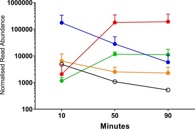
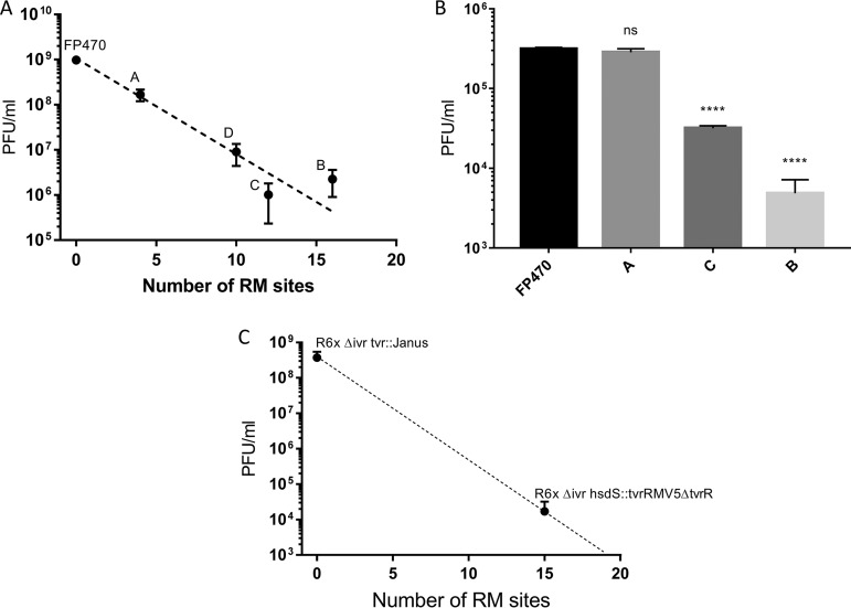
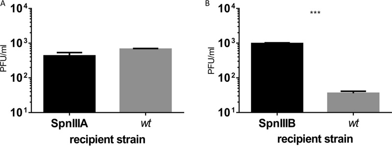
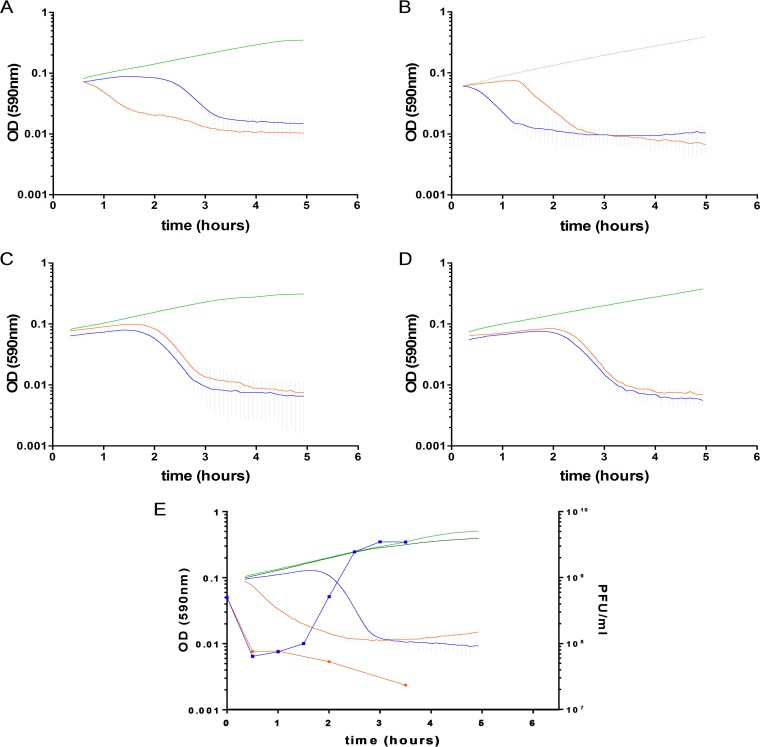
Virus-host interactions are regulated by complex coevolutionary dynamics. In Streptococcus pneumoniae, phase-variable type I restriction-modification (R-M) systems are part of the core genome. We hypothesized that the ability of the R-M systems to switch between six target DNA specificities also has a key role in preventing the spread of bacteriophages. Using the streptococcal temperate bacteriophage SpSL1, we show that the variants of both the SpnIII and SpnIV R-M systems are able to restrict invading bacteriophage with an efficiency approximately proportional to the number of target sites in the bacteriophage genome. In addition to restriction of lytic replication, SpnIII also led to abortive infection in the majority of host cells. During lytic infection, transcriptional analysis found evidence of phage-host interaction through the strong upregulation of the nrdR nucleotide biosynthesis regulon. During lysogeny, the phage had less of an effect on host gene regulation. This research demonstrates a novel combined bacteriophage restriction and abortive infection mechanism, highlighting the importance that the phase-variable type I R-M systems have in the multifunctional defense against bacteriophage infection in the respiratory pathogen S. pneumoniaeIMPORTANCE With antimicrobial drug resistance becoming an increasing burden on human health, much attention has been focused on the potential use of bacteriophages and their enzymes as therapeutics. However, the investigations into the physiology of the complex interactions of bacteriophages with their hosts have attracted far less attention, in comparison. This work describes the molecular characterization of the infectious cycle of a bacteriophage in the important human pathogen Streptococcus pneumoniae and explores the intricate relationship between phase-variable host defense mechanisms and the virus. This is the first report showing how a phase-variable type I restriction-modification system is involved in bacteriophage restriction while it also provides an additional level of infection control through abortive infection.

Keywords: DNA methylation; Streptococcus pneumoniae; abortive infection; bacteriophage genetics; phase variation; restriction-modification system.


Figures

FIG 1
FIG 1
The SpSL1 bacteriophage and spnIII restriction system. (A) The SpSL1 genome displayed in the viral conformation with the cohesive ends flanking the sequence. The color scheme indicates the operon structure, with operon 1 (cds1 to cds4) in orange, operon 2 (cds5 to cds26) in blue, operon 3 (cds27 to cds29) in green, operon 4 (cds30 to cds49) in red, and operon 5 (cds50) in white (GenBank accession number KM882824). (B) The phase-variable type I restriction-modification system spnIII containing the hsdR, hsdM, and variable hsdS genes, in addition to the site-specific recombinase gene creX, with a schematic representation of the recombination. The hsdS gene encodes N-terminal and C-terminal target recognition domains (TRDs). The two N-terminal TRDs are in dark blue and light blue, while the C-terminal TRDs are in red, orange, and purple. The inverted repeats are shown as gray dotted rectangles. The six different hsdS variants, indicated A to F, are shown below (20). (C) A cartoon for a population-based bacteriophage defense that arises from a phase-variable restriction-modification system, where the bacterial genome is methylated in a specific pattern (shown by the colored M on the black line). The bacteriophage would be able to infect and replicate in only one variant (blue), while it would be unable to infect the other variants with different methylation patterns (orange, red, purple, and green) (21).
FIG 2
FIG 2
SpSL1 phage relative gene expression in lytic and lysogenic stages. (A to C) A time course of lytic infection of an spnIII-deleted strain at an MOI of 0.2 at 10 min (A) (green), 50 min (B) (blue), and 90 min (C) (red) after challenge. (D) Gene expression in the lysogen (black). All data are shown in terms of the normalized read coverage. (E) RNA sequencing reads were mapped to the viral conformation of the SpSL1 phage deposited in GenBank (accession number KM882824), even in the case of the lysogen (D). RNA-seq mapping and upper quartile normalization were performed using Rockhopper software. Data were visualized on BAMviewer in the Artemis tool, with the maximum number of reads being 2,000 (small label on the right of each panel). RNA-seq data were deposited at Gene Expression Omnibus GEO (accession number GSE132611).
FIG 3
FIG 3
Bacteriophage SpSL1 gene expression during the lytic cycle. RNA-seq data showing the expression of phage SpSL1 at 10, 50, and 90 min postinfection of spnIII-negative strain FP470. The transcriptional units are numbered 1 to 5, as shown in Fig. 1A. The three early transcriptional units are operon 1 (cds1 to cds4) in orange, operon 2 (cds5 to cds26) in blue, and operon 5 (cds50) in white. The two late transcriptional units are operon 3 (cds27 to cds29) in green and operon 4 (cds30 to cds49) in red.
FIG 4
FIG 4
S. pneumoniae genes are upregulated in response to SpSL1 infection. RNA-seq analysis during SpSL1 infection revealed the upregulation of three S. pneumoniae transcripts preceded by an NrdR binding site and encoding the products of the anaerobic ribonucleoside triphosphate reductase operon (circles; SPD_0187 to SPD_0191, a five-gene operon), the ribonucleoside diphosphate reductase operon (squares; SPD_1041 to SPD_1043, a three-gene operon), and a hypothetical operon encoding an unknown transcriptional regulator and a conserved hypothetical protein (triangles; SPD_1594 and SPD_1595, a two-gene operon). The RNA-seq data were normalized by upper quartile gene normalization and compared with those for a noninfected control to determine the fold change in expression.
FIG 5
FIG 5
Restriction of SpSL1 by phase-variable SpnIII and SpnIV R-M systems. (A) Plaque assays results obtained using SpnDP1004III-unmethylated SpSL1 phage to infect spnDP1004A- to spnDP1004D-locked strains that express single locked copies of one of the hsdS alleles, with FP470 used as a control. (B) The differences observed between the control strain deleted for spnDP1004III (zero sites recognized) and the other mutants were statistically significant for SpSL1. ****, P < 0.001 by a one-way analysis of variance (ANOVA) multiple-comparison test; ns, not significant. (C) The restriction of infection of the spnDP1004III-deleted strain and the spnDP1004IIIA-locked mutant with SpnDP1004IIIA-methylated SpSL1 was not statistically significant (one-way ANOVA multiple-comparison test, P > 0.05), whereas the restriction of infection with the spnDP1004IIIC and spnDP1004IIIB mutants was (one-way ANOVA multiple-comparison test, P < 0.001) (B). Plaque assay results using SpSL1 to test the phase-variable SpnIV system showed differences between the SpnIV-knockout strain and the SpnIV R6x Δivr hsdS::tvrRMV5 ΔtvR recombinant (37) (Student's t test, P < 0.001).
FIG 6
FIG 6
Phase-variable restriction of SpSL1 by SpnIII in a wild-type population. Plaque assays of a wt strain and spnDP1004III-locked mutants. The wt DP1004 strain, harboring 85% and 12% spnDP1004IIIA- and spnDP1004IIIB-positive cells, respectively, was tested for infection by SpnIIIA-methylated phage (A) and SpnIIIB-methylated phage (B) and compared to infection of the spnDP1004IIIA-locked and spnDP1004IIIB-locked strains. (A) Equally efficient SpnIIIA-methylated phage infection of a wt host with a predominance of SpnIIIA cells and of an spnDP1004III-locked mutant. (B) Plaque generation of SpnIIIB-methylated phage. Unrestricted plaque formation in spnDP1004IIIB cells yielded SpnIIIB-methylated phage (data of phage methylation status not shown), while infection of the wt containing 85% spnDP1004IIIA cells (gray bar in panel B) yielded fewer plaques (two-tailed t test, P < 0.001), and all phages obtained were SpnIIIA methylated (data not shown).
FIG 7
FIG 7
Abortive infection by the SpnIII system. Bacterial cell fate and viability were determined by the hsdS allele conformation and by the infecting phage genome methylation status. (A) The spnDP1004IIIA-locked mutant was infected with SpnIIIA-methylated (blue; MOI = 2.5) and SpnIIIA-nonmethylated (orange; MOI = 2.5) SpSL1 phages. (B) The spnDP1004IIIB-locked mutant was infected with SpnIIIB-methylated (orange; MOI = 2.5) and SpnIIIB-nonmethylated (blue; MOI = 2.5) SpSL1 phages. In both cases, the nonrestricted phage killed the cells after completion of the lytic cycle, while the supposedly restricted phage induced a rapid and progressive lysis. When infecting an spnDP1004III deletion mutant (C), the phage underwent a lytic cycle irrespective of its methylation status (SpnIIIA methylated, blue; SpnIIIB methylated, orange; MOI = 2.5). The same outcome was achieved by inactivating the restriction subunit of spnDP1004III alone (D). In panels A to E, uninfected bacterial strains are depicted in green. The one-step growth curves (infecting free viral particles were measured each 30 min after infection) in panel E confirmed the production of phage progeny when SpSL1 was not restricted (as with the SpnIIIA-methylated phage infecting the spnDP1004IIIA-locked mutant [blue lines]; light green, uninfected control) or the absence of phage replication when SpSL1 was restricted by SpnDP1004III (as with the SpnIIIA-methylated phage infecting the spnDP1004IIIB-locked mutant [orange lines]; dark green, uninfected control). The SpSL1 burst size was 20 PFU.
FIG 8
FIG 8
McrBC and LytA are not responsible for abortive infection. The SpnMcrBC type IV R-M system and the autolysin LytA have no effect on the Abi phenotype. Mutants for mcrBC (A) and lytA (B) were constructed in an spnDP1004IIIA-locked background, and the pairs of recombinant strains were infected with SpnIIIA-methylated (blue; MOI = 2.5) and SpnIIIA-unmethylated (orange; MOI = 2.5) SpSL1 phage. Uninfected controls are shown in green. Both mcrBC and lytA mutants showed nearly immediate lysis upon SpnIIIA-unmethylated phage infection, indicative of an unmodified Abi phenotype.

Similar articles

Cited by

References

    1. Clokie MR, Millard AD, Letarov AV, Heaphy S. 2011. Phages in nature. Bacteriophage 1:31–45. doi:10.4161/bact.1.1.14942. - DOI - PMC - PubMed
    1. Koskella B, Brockhurst MA. 2014. Bacteria-phage coevolution as a driver of ecological and evolutionary processes in microbial communities. FEMS Microbiol Rev 38:916–931. doi:10.1111/1574-6976.12072. - DOI - PMC - PubMed
    1. Stern A, Sorek R. 2011. The phage-host arms race: shaping the evolution of microbes. Bioessays 33:43–51. doi:10.1002/bies.201000071. - DOI - PMC - PubMed
    1. Ercoli G, Fernandes VE, Chung WY, Wanford JJ, Thomson S, Bayliss CD, Straatman K, Crocker PR, Dennison A, Martinez-Pomares L, Andrew PW, Moxon ER, Oggioni MR. 2018. Intracellular replication of Streptococcus pneumoniae inside splenic macrophages serves as a reservoir for septicaemia. Nat Microbiol 3:600–610. doi:10.1038/s41564-018-0147-1. - DOI - PMC - PubMed
    1. Kadioglu A, Weiser JN, Paton JC, Andrew PW. 2008. The role of Streptococcus pneumoniae virulence factors in host respiratory colonization and disease. Nat Rev Microbiol 6:288–301. doi:10.1038/nrmicro1871. - DOI - PubMed

Publication types