. 2019 Jul 10;8(7):bio044529.
doi: 10.1242/bio.044529.

Analysis of novel hyperosmotic shock response suggests 'beads in liquid' cytosol structure

Affiliations

Analysis of novel hyperosmotic shock response suggests 'beads in liquid' cytosol structure

Alexander I Alexandrov et al. Biol Open. .

Abstract

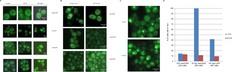
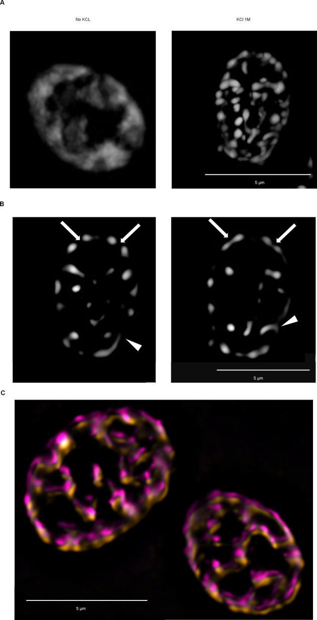
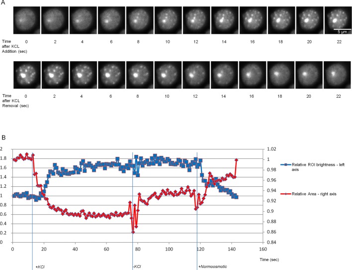
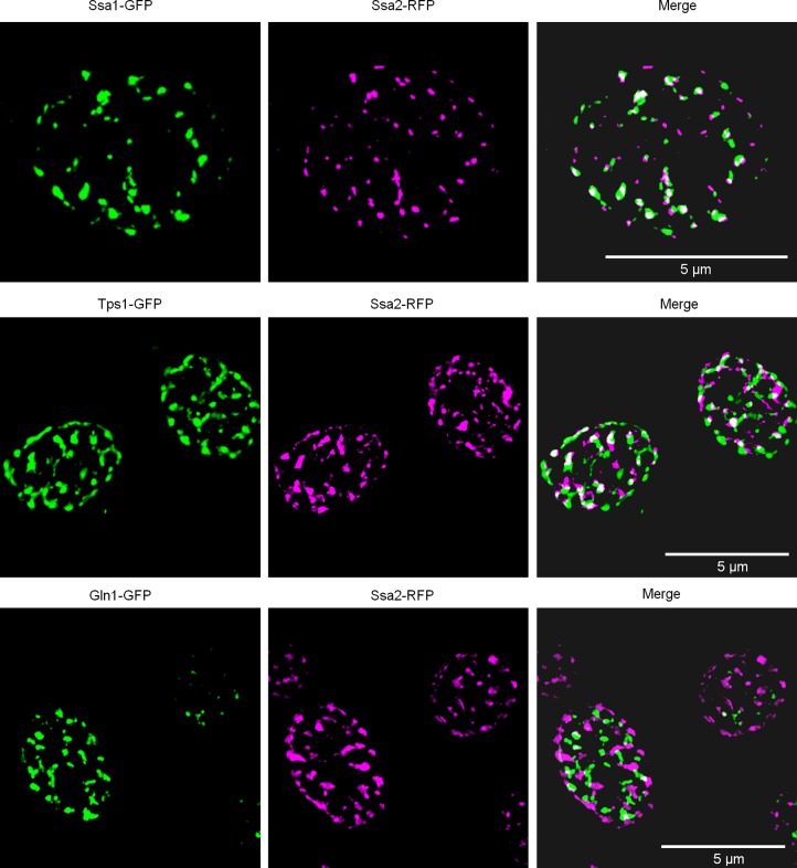
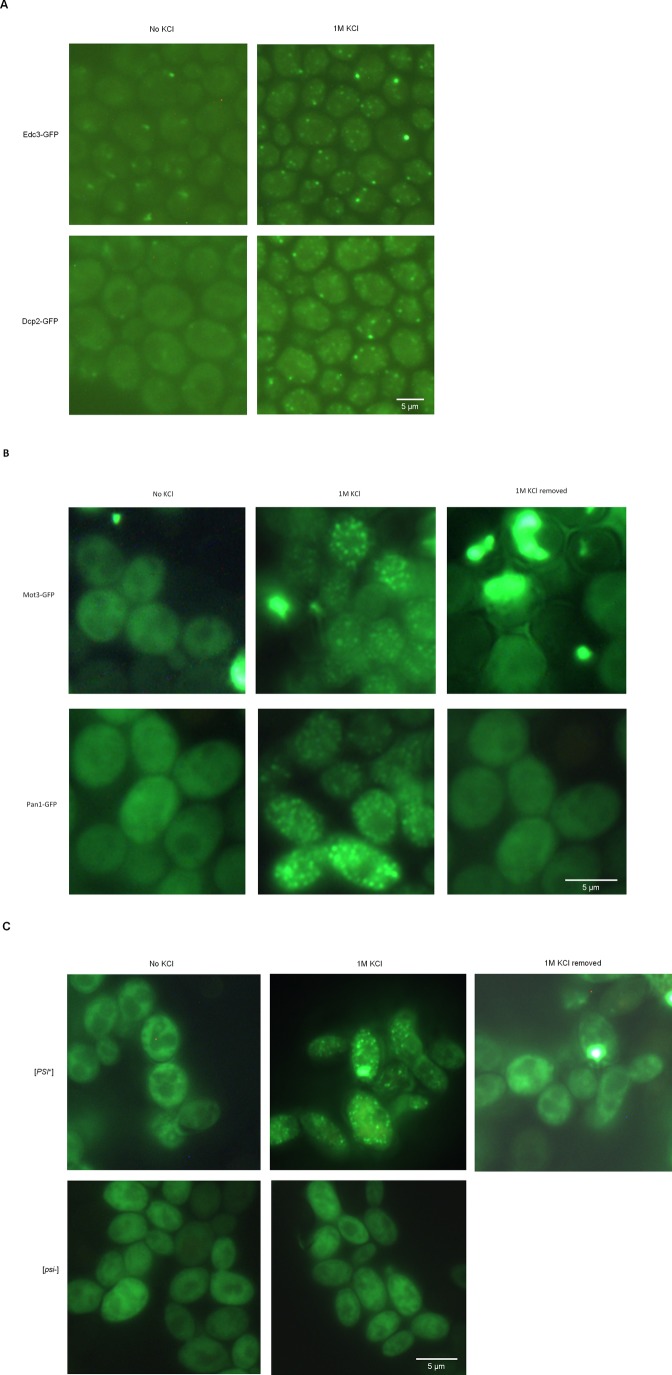
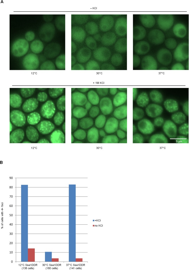
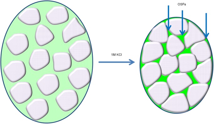
Proteins can aggregate in response to stresses, including hyperosmotic shock. Formation and disassembly of aggregates is a relatively slow process. We describe a novel instant response of the cell to hyperosmosis, during which chaperones and other proteins form numerous foci with properties uncharacteristic of classical aggregates. These foci appeared/disappeared seconds after shock onset/removal, in close correlation with cell volume changes. Genome-wide and targeted testing revealed chaperones, metabolic enzymes, P-body components and amyloidogenic proteins in the foci. Most of these proteins can form large assemblies and for some, the assembled state was pre-requisite for participation in foci. A genome-wide screen failed to identify genes whose absence prevented foci participation by Hsp70. Shapes of and interconnections between foci, revealed by super-resolution microscopy, indicated that the foci were compressed between other entities. Based on our findings, we suggest a new model of cytosol architecture as a collection of numerous gel-like regions suspended in a liquid network. This network is reduced in volume in response to hyperosmosis and forms small pockets between the gel-like regions.

Keywords: Aggregation; Amyloid; Chaperone; Cytoplasm; Foci; Hyperosmotic shock; Liquid–liquid phase separation; P-bodies; Yeast.


Conflict of interest statement

Competing interestsThe authors declare no competing or financial interests.

Figures

Fig. 1.
Fig. 1.
Chaperones can form OSFs under various stress conditions both in S. cerevisiae and O. parapolymorpha. (A) Cells of the BY4741 strain bearing the indicated GFP-fusion protein were grown in YP-Gly medium and then transferred onto medium with the indicated concentration of KCl, glycerol or sorbitol. (B) Cells bearing the indicated GFP-fusion chaperone were grown in YP-Gly or YPD medium to logarithmic phase and transferred onto the same medium supplemented with 1M KCl. (C) Cells of O. parapolymorpha, producing the closest Ssa1 homologue tagged with GFP (see Materials and Methods) were grown in YP-Gly medium to logarithmic phase and transferred onto YPD medium with 1M KCl. (D) Analysis of foci numbers in populations of cells used in B and C, with the vertical axis depicting the percentage of cells containing more than three foci. OP, O. parapolymorpha cells. Scale bars: 5 µm.
Fig. 2.
Fig. 2.
SIM reveals bridges between OSFs as well as small OSF protuberances. Cells producing the Ssa1-DDR protein were collected in the logarithmic phase of growth from YP-Gly medium, and either subjected or not subjected to hyperosmotic shock with 1M KCl in the same medium. (A) Maximum intensity projection of a representative shocked and non-shocked cell. (B) Selected optic sections from the same shocked cell shown in A that contain OSFs with protuberances or interconnections between adjacent OSFs. Arrows indicate interconnections, while protuberances are indicated by arrowheads. (C) Two SIM images (average intensity projections) of the same cells taken within a 2-min interval (initial image pseducolored magenta, with orange after 2 min). An offset was introduced in order to facilitate viewing of changes in configuration of features. Scale bars: 5 µm.
Fig. 3.
Fig. 3.
Dynamics of Ssa1-GFP OSF formation and disappearance. (A) Time-lapse images of OSF formation and disappearance in response to onset and removal of hyperosmosis, respectively, obtained using cells expressing the Ssa1-GFP protein. Images were obtained using confocal real-time microscopy. Scale bar: 5 µm. (B) Time course of changes in cytoplasmic area and OSF formation/disappearance. Vertical lines depict the time points at which the KCl-induced hyperosmotic shock was administered (+KCl), the time at which medium containing KCl was aspirated (−KCl), as well as the time at which normoosmotic medium was added (+Normoosmotic). The presented graph was obtained from the cell images in A, except with higher temporal resolution. Ten other cells were also used to construct graph with similar results. The Ssa1-GFP protein was used instead of Ssa1-DDR to provide a stronger signal without photobleaching. The large aggregate that is constant through both time courses is an IPOD-like inclusion of Ssa1-GFP that has no relation to OSFs. Fluctuation in the graph during KCl removal and slight change of apparent cell area afterwards is due to vibrations of the sample during solution aspiration, which affected microscope focusing.
Fig. 4.
Fig. 4.
OSF-forming proteins show only partial or absent colocalization. Diploid cells expressing pairs of the indicated GFP/tagRFP-tagged proteins were grown to mid-log phase in YP-Gly medium, transferred onto the same medium with 1M KCl and visualized by SIM microscopy. Scale bars: 5 µm.
Fig. 5.
Fig. 5.
P-body proteins and proteins with amyloidogenic domains can form OSFs. (A) Cells producing the indicated GFP-fusion proteins were grown in YP-Gly medium and washed in SC-Gly medium to reduce background medium fluorescence. The cells were subsequently subjected to hyperosmotic shock (1M KCl) in SC-Gly. (B) Cells of the 74D-694 [psi] strain harboring the plasmids expressing PrDMot3-GFP or PrDPan1-GFP were grown in SC-Gal medium to induce GFP-fusion protein production and then transferred onto the same medium supplemented with 1M KCl. (C) Cells of the 74D-694 strain harboring the plasmid expressing PrDSup35-GFP, as well as containing or lacking prion amyloids of Sup35 ([PSI+] or [psi], respectively) were grown in SCGly medium and then transferred onto the same medium with 1M KCl. The growth of cells in SCGly was required to observe diffuse fluorescence of the protein in normo-osmotic conditions, since high production of PrdSup35-GFP upon growth of [PSI+] cells in SC-Gal resulted in almost complete accumulation of this protein in IPOD-like inclusions. Due to the presence of the ade1-14 mutation, additional adenine was added to the medium to reduce of the amount of autofluorescent red pigment accumulated in cells of the ade1 mutants. Scale bars: 5 µm.
Fig. 6.
Fig. 6.
Conditions of mild stress during growth stimulate formation of OSFs by Ssa1-DDR. (A) Cells of the BY4741 strain bearing the Ssa1-DDR fusion protein were grown in YPD under the indicated conditions and then subjected to hyperosmotic shock. (B) Analysis of foci numbers in populations of cells used in A, with the y-axis depicting the percentage of cells containing 4+ foci. Exposition times were adjusted to have approximately equal intensity in the various samples. Scale bar: 5 µm.
Fig. 7.
Fig. 7.
Schematic representation of the proposed mechanism of OSF emergence during hyperosmotic shock. In response to hyperosmosis, the cell loses water, concentrating the liquid part of the cytoplasm (green). Putative solid components bunch together, forming concentrated pockets of liquid (more vivid green), that we propose to correspond to OSFs. Nature of solid components may vary, including membrane organelles in part, but also unknown cytosolic structures.

References

    1. Agaphonov M. O. (2017). Improvement of a yeast self-excising integrative vector by prevention of expression leakage of the intronated Cre recombinase gene during plasmid maintenance in Escherichia coli. FEMS Microbiol. Lett. 364, fnx222 10.1093/femsle/fnx222 - DOI - PubMed
    1. Agaphonov M., Romanova N., Choi E.-S. and Ter-Avanesyan M. (2009). A novel kanamycin/G418 resistance marker for direct selection of transformants in Escherichia coli and different yeast species. Yeast 27, 189-195. 10.1002/yea.1741 - DOI - PubMed
    1. Alberti S., Halfmann R., King O., Kapila A. and Lindquist S. (2009). A systematic survey identifies prions and illuminates sequence features of prionogenic proteins. Cell 137, 146-158. 10.1016/j.cell.2009.02.044 - DOI - PMC - PubMed
    1. Alexandrov A. I. and Dergalev A. A. (2019). Increasing throughput of manual microscopy of cell suspensions using solid medium pads. MethodsX 6, 329-332. 10.1016/j.mex.2019.02.010 - DOI - PMC - PubMed
    1. Bracha D., Walls M. T., Wei M.-T., Zhu L., Kurian M., Avalos J. L., Toettcher J. E. and Brangwynne C. P. (2018). Mapping local and global liquid phase behavior in living cells using photo-oligomerizable seeds. Cell 175, 1467-1480.e13. 10.1016/j.cell.2018.10.048 - DOI - PMC - PubMed