. 2019 Aug 1;8(8):bio044370.
doi: 10.1242/bio.044370.

Reduced mTORC1-signalling in retinal progenitor cells leads to visual pathway dysfunction

Affiliations

Reduced mTORC1-signalling in retinal progenitor cells leads to visual pathway dysfunction

Iwan Jones et al. Biol Open. .

Abstract

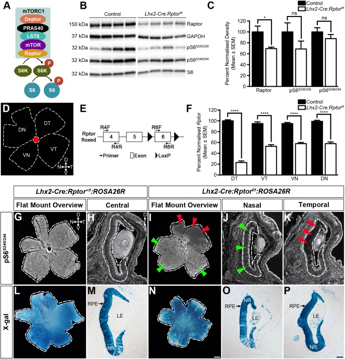
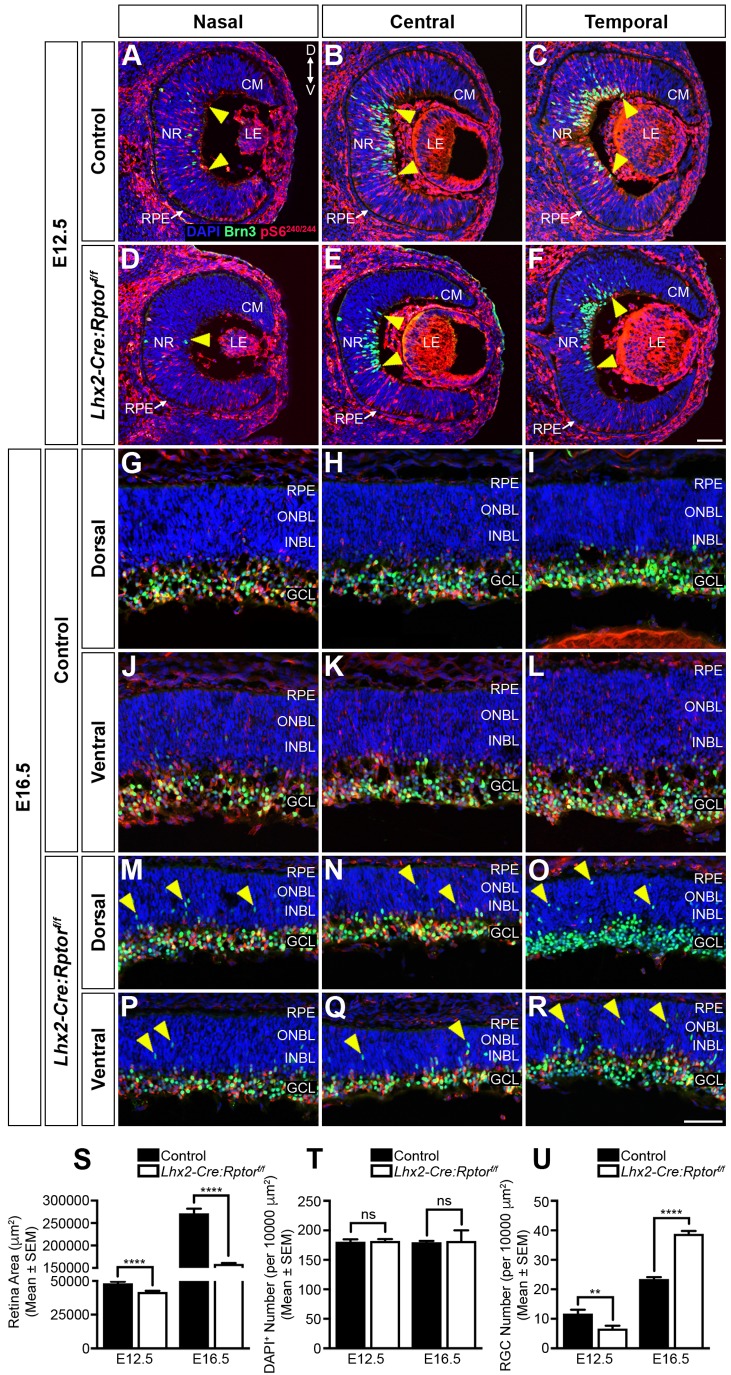
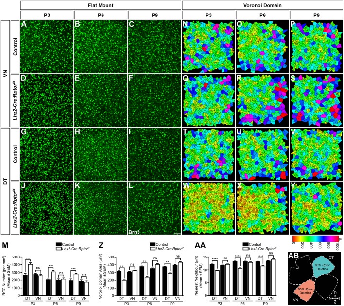
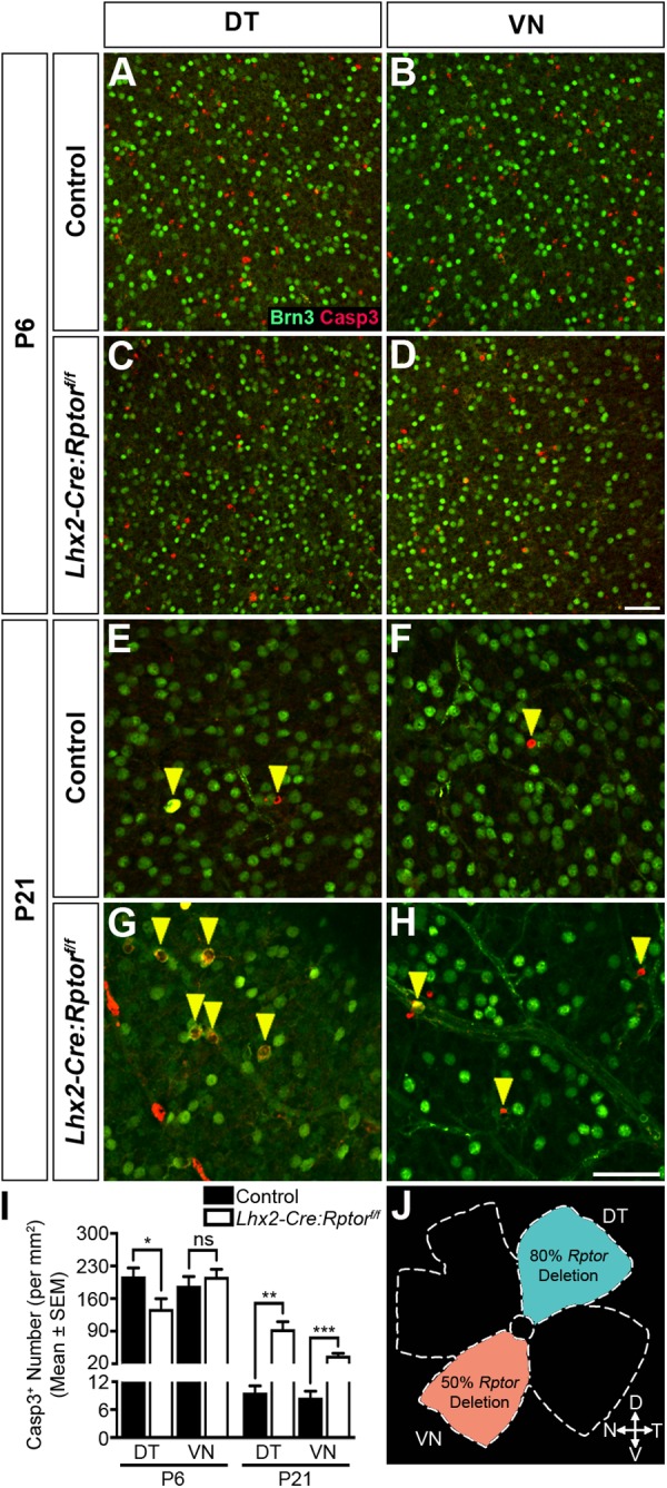
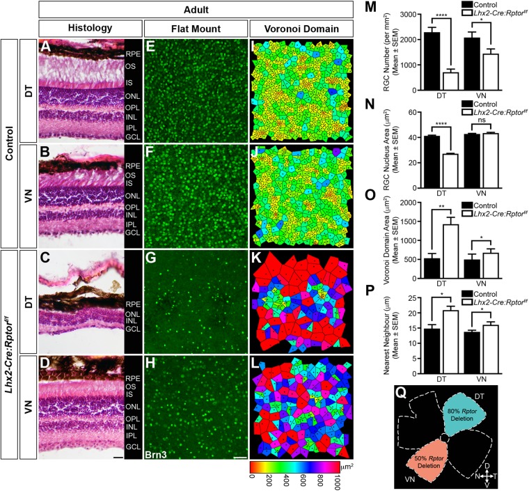
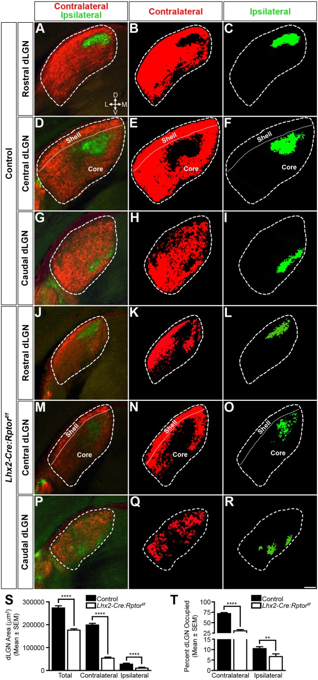
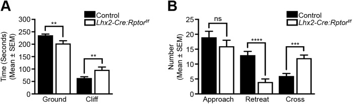
Development of the vertebrate central nervous system involves the co-ordinated differentiation of progenitor cells and the establishment of functional neural networks. This neurogenic process is driven by both intracellular and extracellular cues that converge on the mammalian target of rapamycin complex 1 (mTORC1). Here we demonstrate that mTORC1-signalling mediates multi-faceted roles during central nervous system development using the mouse retina as a model system. Downregulation of mTORC1-signalling in retinal progenitor cells by conditional ablation of Rptor leads to proliferation deficits and an over-production of retinal ganglion cells during embryonic development. In contrast, reduced mTORC1-signalling in postnatal animals leads to temporal deviations in programmed cell death and the consequent production of asymmetric retinal ganglion cell mosaics and associated loss of axonal termination topographies in the dorsal lateral geniculate nucleus of adult mice. In combination these developmental defects induce visually mediated behavioural deficits. These collective observations demonstrate that mTORC1-signalling mediates critical roles during visual pathway development and function.

Keywords: RGCs; Raptor; Retina; Visual cliff test; dLGN; mTORC1.


Conflict of interest statement

Competing interestsThe authors declare no competing or financial interests.

Figures

Fig. 1.
Fig. 1.
Conditional deletion of Rptor leads to domain-specific reductions in mTORC1-signalling. (A) The mTORC1 is assembled from Deptor, PRAS40, LST8, mTOR and Raptor. This multimeric complex regulates protein translation in response to nutritional and environmental cues through the phosphorylation of the 40S ribosomal protein S6 (pS6). (B) Representative immunoblot analyses of Raptor, GAPDH, pS6S235/236, pS6S240/244 and S6 in retinal extracts harvested from control (n=4) and Lhx2-Cre:Tsc1f/f (n=4) mice at P1. (C) Densitometry analyses of Raptor, pS6S235/236 and pS6S240/244 in retinal extracts harvested from control (n=6) and Lhx2-Cre:Rptorf/f (n=6) mice at P1. Lhx2-Cre:Rptorf/f mice exhibit a significant 30% decrease in Raptor levels compared to littermate controls. No significant differences in pS6S235/236 and pS6S240/244 levels were observed. Raptor densitometry data were normalised against GAPDH while pS6S235/236 and pS6S240/244 values were normalised against S6. (D) Schematic diagram detailing the dissection strategy employed for harvesting retinal quadrants. (E) Schematic diagram detailing the qPCR strategy employed to determine the level of Rptor recombination in the retinal quadrants. The level of PCR product generated by the target primer pair (R6F and R6R) was normalised against that amplified from the internal control primer pair (R4F and R4R). (F) qPCR analysis of normalised Rptor levels in retinae harvested from control (n=6) and Lhx2-Cre:Rptorf/f (n=7) mice at 6 weeks of age. Mutant animals exhibit a domain-specific pattern of Rptor recombination with approximately 80% deletion being observed in the DT retina while the remaining quadrants exhibit close to a 50% deletion rate. (G–K) Representative immunohistochemical staining of retinae harvested from Lhx2-Cre:Rptor+/f:ROSA26R (G–H) and Lhx2-Cre:Rptorf/f:ROSA26R (I–K) mice at P1. Lhx2-Cre:Rptor+/f:ROSA26R animals exhibit widespread pS6S240/244 distribution due to canonical mTORC1-signalling mediated by the remaining wild-type Rptor allele. In contrast, no pS6S240/244 was observed in the DT retina (I,K, red triangles) of Lhx2-Cre:Rptorf/f:ROSA26R mice while modest pS6S240/244 levels were observed within the remaining regions (I,J, green triangles). (L–P) Representative lineage tracing analyses of retinae taken from Lhx2-Cre:Rptor+/f:ROSA26R (L,M) and Lhx2-Cre:Rptorf/f:ROSA26R (N–P) mice at P1. All mice exhibit widespread X-gal staining throughout the retina thus confirming global Lhx2-Cre transgene expression and subsequent Cre-mediated recombination of the ROSA26R allele. Note that radial columns of X-gal mosaicism are observed in both Lhx2-Cre:Rptor+/f:ROSA26R and Lhx2-Cre:Rptorf/f:ROSA26R mice due to variable ROSA26R recombination in the ventral retina (M,O). The broken line demarcates the border of the retina in all images. All data represents the mean±s.e.m. Statistical differences were calculated using unpaired two-tailed Student's t-tests. P-values are denoted as follows: *P≤0.05 and ****P≤0.0001. Scale bars: (G,I,L,N) 500 µm; (H,J,K,M,O,P) 100 µm. Abbreviations: D, dorsal; Deptor, DEP domain containing mTOR interacting protein; DN, dorsonasal; DT, dorsotemporal; GAPDH, glyceraldehyde 3-phosphate dehydrogenase; kDa, kilodalton; LE, lens; LST8, lethal with SEC13 protein 8; mTOR, mechanistic target of rapamycin; mTORC1, mechanistic target of rapamycin complex 1; N, nasal; NR, neural retina; P, postnatal day; PRAS40, proline rich AKT substrate 40 kDa; pS6, phosphorylated ribosomal protein S6; pS6K, phosphorylated ribosomal protein S6 kinase; Raptor, regulatory protein associated with mTOR; RPE, retinal pigment epithelium; S6, ribosomal protein S6; S6K, ribosomal protein S6 kinase; T, temporal; V, ventral; VN, ventronasal; VT, ventrotemporal; X-gal, 5-bromo-4-chloro-3-indolyl-β-D-galactopyranoside.
Fig. 2.
Fig. 2.
Domain-specific reduction in mTORC1-signalling leads to aberrant RGC neurogenesis and radial migration deficits during embryonic development. RGC differentiation and mTORC1-activity was assessed by immunohistochemistry at developmental time points that coincided with neurogenic onset (E12.5) and radial migration (E16.5) milestones. (A–F) Representative coronal sections through the nasal to temporal axis of control (A–C) and mutant (D–F) animals at E12.5 demonstrates the delayed appearance of RGCs and reduced mTORC1-signalling in Lhx2-Cre:Rptorf/f mice. The yellow triangles represent the dorsal and ventral boundaries of the differentiating Brn3+ RGC population, respectively. Note that both control and mutant animals display similar nasallow to temporalhigh RGC distribution gradients. (G–R) Representative coronal sections through the nasal to temporal axis of control (G–L) and Lhx2-Cre:Rptorf/f (M–R) animals at E16.5 reveals ectopic Brn3+ cells (yellow arrowheads) in both the dorsal and ventral neuroblastic layers of mutant mice. Moreover, a reduced level of mTORC1-signalling is observed throughout the whole extent of the retina in Lhx2-Cre:Rptorf/f animals with the DT region (O) exhibiting a complete lack of pS6S240/244 immunoreactivity. (S) Quantitative analysis demonstrates that the area of the retina in Lhx2-Cre:Rptorf/f mice was significantly smaller during both neurogenesis (E12.5) and radial migration (E16.5). (T) Quantitative analysis demonstrates that the density of DAPI+ cells in the retina of both control and Lhx2-Cre:Rptorf/f mice were comparable during both neurogenesis (E12.5) and radial migration (E16.5). (U) Quantitative analysis demonstrates that the density of Brn3+ cells in the retina of Lhx2-Cre:Rptorf/f mice was significantly reduced during neurogenesis (E12.5) but the subsequent over-production of RGCs leads to a significant increase in their numbers during radial migration (E16.5). The number of eyes analysed at each age was as follows: E12.5 (control, n=8; Lhx2-Cre:Rptorf/f, n=8); E16.5 (control, n=8; Lhx2-Cre:Rptorf/f, n=12). All data represents the mean±s.e.m. Statistical differences were calculated using unpaired two-tailed Student's t-tests. P-values are denoted as follows: **P≤0.01 and ****P≤0.0001. Scale bar: (A–F) 50 µm; (G–R) 50 µm. Abbreviations: CM, ciliary margin; D, dorsal; GCL, ganglion cell layer; INBL, inner neuroblastic layer; LE, lens; NR, neural retina; ONBL, outer neuroblastic layer; RPE, retinal pigment epithelium; V, ventral.
Fig. 3.
Fig. 3.
Domain-specific reduction in mTORC1-signalling leads to an over-production of RGCs in the dorsotemporal retina during postnatal development. Retinae were harvested from control and Lhx2-Cre:Rptorf/f mice during postnatal development to determine the mosaic arrangement of RGCs in the VN and DT regions (AB). (A–L) Representative flat-mount analyses of the VN and DT regions of control and Lhx2-Cre:Rptorf/f mice at P3 (A,D,G,J), P6 (B,E,H,K) and P9 (C,F,I,L) demonstrate that both domains in wild-type animals in addition to the VN region of Lhx2-Cre:Rptorf/f mice (A–I) exhibit a comparable number of Brn3+ cells that are dispersed across the surface of the retina in similar arrays. In contrast, a seemingly greater number of RGCs are present in the DT region of mutant animals at all ages analysed (J–L). (M) Quantification of RGC number in the DT and VN quadrants of control and Lhx2-Cre:Rptorf/f mice at P3, P6 and P9. Mutant mice have a significant increase in the mean number of Brn3+ cells within the DT region at all ages analysed. (N–Y) The regularity of RGC mosaics in the VN and DT regions of control and Lhx2-Cre:Rptorf/f mice at P3 (N,Q,T,W), P6 (O,R,U,X) and P9 (P,S,V,Y) was quantified by Voronoi domain and nearest neighbour analyses. Representative heat map diagrams reveal comparable RGC territories within both domains of wild-type animals in addition to the VN region of Lhx2-Cre:Rptorf/f mice (N–V). In contrast, the increased number of RGCs in the DT domain of Lhx2-Cre:Rptorf/f mice (M) resulted in smaller Voronoi domain areas at all ages analysed (W–Y). (Z,AA) Quantitative analyses of the Voronoi domain (Z) and nearest neighbour distances (AA) demonstrate that RGCs solely within the DT region of Lhx2-Cre:Rptorf/f mice exhibit significantly reduced Voronoi domain areas and nearest neighbour distances compared to control littermates due to the increased number of cells in this quadrant (M). The corresponding Voronoi domain and nearest neighbour distance histogram plots are presented in Figs S8S10. (AB) Schematic diagram detailing the retinal quadrants imaged for the Voronoi domain and nearest neighbour analyses. The number of retinae analysed at each age was as follows: P3 (control, n=6; Lhx2-Cre:Rptorf/f, n=6); P6 (control, n=4; Lhx2-Cre:Rptorf/f, n=4); P9 (control, n=11; Lhx2-Cre:Rptorf/f, n=8). All data represents the mean±s.e.m. Statistical differences were calculated using unpaired two-tailed Student's t-tests. P-values are denoted as follows: **P≤0.01, ***P≤0.001 and ****P≤0.0001. Scale bar: (A–L) 50 µm. Abbreviations: D, dorsal; DT, dorsotemporal; N, nasal; P, postnatal day; T, temporal; V, ventral; VN, ventronasal.
Fig. 4.
Fig. 4.
Domain-specific reduction in mTORC1-signalling leads to aberrant RGC apoptosis during postnatal development. Retinae were harvested from control and Lhx2-Cre:Rptorf/f mice during postnatal development to determine the number of apoptotic cells in the DT and VN regions (J). (A–D) Representative flat-mount images of the DT and VN retina in control and Lhx2-Cre:Rptorf/f mice at P6 demonstrate that both domains in wild-type animals (A–B) in addition to the VN region of Lhx2-Cre:Rptorf/f (D) mice exhibit a comparable number of Casp3+ cells. In contrast, a decreased number of apoptotic cells are observed in the DT region of mutant animals (C). (E–H) Representative flat-mount analyses of the DT and VN retina in control and Lhx2-Cre:Rptorf/f mice at P21 demonstrate that both domains in wild-type animals (E,F) exhibit solitary Casp3+ cells (yellow arrowheads). In contrast, an increased number of apoptotic cells (yellow arrowheads) was observed in both the DT (G) and VN (H) regions of Lhx2-Cre:Rptorf/f animals. (I) Quantification of Casp3+ cell number in the DT and VN quadrants of control and Lhx2-Cre:Rptorf/f mice. Mutant mice have a significant decrease in the mean number of apoptotic cells within the DT region at P6. Contrastingly, mutant mice have a significant increase in the mean number of apoptotic cells in both the DT and VN region at P21. (J) Schematic diagram detailing the retinal quadrants imaged for the apoptosis analyses. The number of retina analysed at each age was as follows: P6 (control, n=12; Lhx2-Cre:Rptorf/f, n=10); P21 (control, n=6; Lhx2-Cre:Rptorf/f, n=14). All data represents the mean±s.e.m. Statistical differences were calculated using unpaired two-tailed Student's t-tests. P-values are denoted as follows: *P≤0.05, **P≤0.01 and ***P≤0.001. Scale bar: (A–D) 50 µm; (E–H) 50 µm. Abbreviations: D, dorsal; DT, dorsotemporal; N, nasal; P, postnatal day; T, temporal; V, ventral; VN, ventronasal.
Fig. 5.
Fig. 5.
Domain-specific reduction in mTORC1-signalling leads to aberrant retinal morphology and disorganised RGC mosaics in adult mice. Enucleated eyes were harvested from control (n=8) and Lhx2-Cre:Rptorf/f mice (n=8) at 7 weeks of age and the morphological appearance of the retina in addition to the mosaic arrangement of RGCs in the DT and VN regions (Q) was determined. (A–D) Representative coronal eye sections taken from control mice (A,B) demonstrates that the retina in both the DT and VN regions exhibits a characteristic laminated structure with three nuclear layers (ONL, INL and GCL) being interconnected by two plexiform layers (OPL and IPL). In contrast, while the morphology of the retina in Lhx2-Cre:Rptorf/f mice (C,D) also displays comparable stratification, the apicobasal thickness of the DT region is reduced due to a decrease in the number of cells within the nuclear layers (C) while both regions exhibit a disorganised GCL. (E–H) Representative flat-mount images of control retinae demonstrate that the RGCs in both the DT (E) and VN (F) quadrants are dispersed in well-ordered arrays. In contrast, the Brn3+ cells in the DT (G) and VN regions (H) of Lhx2-Cre:Rptorf/f animals appear fewer in number with asymmetric distribution. (I–L) The spatial properties of the RGC mosaics were determined by Voronoi domain and nearest neighbour analyses. Representative heat map diagrams reveal greater variability in Voronoi domain areas and nearest neighbour distances within Lhx2-Cre:Rptorf/f animals (K,L) compared to control littermates (I,J). (M) Quantification of RGC number in the DT and VN quadrants of control and Lhx2-Cre:Rptorf/f mice. Mutant mice have a significant decrease in the mean number of Brn3+ cells in both domains. (N) Quantification of RGC nucleus area in the DT and VN quadrants of control and Lhx2-Cre:Rptorf/f mice. Mutant mice have a significant decrease in nucleus area in the DT region. (O,P) Quantitative analyses of the Voronoi domain (O) and nearest neighbour distances (P) demonstrate that RGCs within the DT and VN regions of Lhx2-Cre:Rptorf/f mice exhibit significantly larger domain areas and nearest neighbour distances compared to control littermates. The corresponding Voronoi domain and nearest neighbour distance histogram plots are presented in Fig. S12. (Q) Schematic diagram detailing the retinal quadrants imaged for the Voronoi domain and nearest neighbour analyses. All data represents the mean±s.e.m. Statistical differences were calculated using unpaired two-tailed Student's t-tests. P-values are denoted as follows: *P≤0.05, **P≤0.01 and ****P≤0.0001. Scale bar: (A–D) 100 µm; (E–H) 50 µm. Abbreviations: D, dorsal; DT, dorsotemporal; GCL, ganglion cell layer; INL, inner nuclear layer; IPL, inner plexiform layer; IS, inner segments; N, nasal; ONL, outer nuclear layer; OPL, outer plexiform layer; OS, outer segments; RPE, retinal pigment epithelium; T, temporal; V, ventral; VN, ventronasal.
Fig. 6.
Fig. 6.
Aberrant RGC mosaics induce loss of retinogeniculate termination topographies in adult mice. Binocular projection patterns in the dLGN of control (n=8) and Lhx2-Cre:Rptorf/f (n=8) mice were visualised by intraocular injections of fluorescently labelled CTB at 6 weeks of age. (A–I) Representative coronal sections through the dLGN of control animals to visualise the termination topographies in rostral (A–C), central (D–F) and caudal (G–I) positions. Binocular projections (A,D,G) were well segregated into distinct contralateral (B,E,H) and ipsilateral inputs (C,F,I). (J–R) Representative coronal sections through the dLGN of mutant animals to show the retinogeniculate topographies in the rostral (J–L), central (M–O) and caudal (P–R) positions. The total dLGN area was reduced (J,M,P) and large unoccupied contralateral territories (K,N,Q) were observed. In addition, the termination area of the ipsilateral projection was also reduced in size and presented as clusters of distinct arbors (L,O,R). The contralateral and ipsilateral panels are presented as binarised images to allow for better visualisation of the termination topographies. Dashed lines define the border of the dLGN in all images. Dotted lines define the division between the shell and core domains in the central dLGN images (S). Quantitative area analysis demonstrated that the total dLGN and contributing contralateral and ipsilateral termination areas were significantly reduced in Lhx2-Cre:Rptorf/f mice when compared to control animals. (T) Quantitative occupation analysis demonstrated that the percentage of dLGN area occupied by the contralateral and ipsilateral projections was significantly reduced in Lhx2-Cre:Rptorf/f mice. All data represents the mean±s.e.m. Statistical differences were determined using unpaired two-tailed Student's t-tests. P-values are denoted as follows: **P≤0.01 and ****P≤0.0001. Scale bar: 100 µm. Abbreviations: D, dorsal; dLGN, dorsal lateral geniculate nucleus; L, lateral; M, medial; T, temporal; V, ventral.
Fig. 7.
Fig. 7.
The cumulative effects of Rptor-ablation lead to visual behaviour deficits. Visual cliff analyses of control (n=20) and Lhx2-Cre:Rptorf/f (n=13) mice between 6 and 10 weeks of age. (A) Visual cliff data analysed for total time spent on either the ground or cliff side of the testing arena. Lhx2-Cre:Rptorf/f mice spend significantly less time on the ground and consequently more time on the cliff side. (B) Visual cliff data analysed for total number of (i) approaches to the central beam, (ii) retreats to the ground side following an approach and (iii) crosses to the cliff side following an approach. Both groups made a comparable number of approaches towards the central beam but Lhx2-Cre:Rptorf/f mice made significantly more crosses onto the cliff side of the testing arena with a consequent reduction in the number of retreats. All data represents the mean±s.e.m. Statistical differences were calculated using unpaired two-tailed Student's t-tests. P-values are denoted as follows: **P≤0.01, ***P≤0.001 and ****P≤0.0001.

Similar articles

Cited by

References

    1. Agathocleous M. and Harris W. A. (2009). From progenitors to differentiated cells in the vertebrate retina. Annu. Rev. Cell Dev. Biol. 25, 45-69. 10.1146/annurev.cellbio.042308.113259 - DOI - PubMed
    1. Amini R., Rocha-Martins M. and Norden C. (2017). Neuronal migration and lamination in the vertebrate retina. Front. Neurosci. 11, 742 10.3389/fnins.2017.00742 - DOI - PMC - PubMed
    1. Bansal A., Singer J. H., Hwang B. J., Xu W., Beaudet A. and Feller M. B. (2000). Mice lacking specific nicotinic acetylcholine receptor subunits exhibit dramatically altered spontaneous activity patterns and reveal a limited role for retinal waves in forming ON and OFF circuits in the inner retina. J. Neurosci. 20, 7672-7681. 10.1523/JNEUROSCI.20-20-07672.2000 - DOI - PMC - PubMed
    1. Bassett E. A. and Wallace V. A. (2012). Cell fate determination in the vertebrate retina. Trends Neurosci. 35, 565-573. 10.1016/j.tins.2012.05.004 - DOI - PubMed
    1. Bateman J. M. and McNeill H. (2004). Temporal control of differentiation by the insulin receptor/tor pathway in Drosophila. Cell 119, 87-96. 10.1016/j.cell.2004.08.028 - DOI - PubMed