. 2019 Jul 23;116(30):15236-15243.
doi: 10.1073/pnas.1905705116. Epub 2019 Jul 8.

Trpc5 deficiency causes hypoprolactinemia and altered function of oscillatory dopamine neurons in the arcuate nucleus

Affiliations

Trpc5 deficiency causes hypoprolactinemia and altered function of oscillatory dopamine neurons in the arcuate nucleus

Thomas Blum et al. Proc Natl Acad Sci U S A. .

Abstract

Dopamine neurons of the hypothalamic arcuate nucleus (ARC) tonically inhibit the release of the protein hormone prolactin from lactotropic cells in the anterior pituitary gland and thus play a central role in prolactin homeostasis of the body. Prolactin, in turn, orchestrates numerous important biological functions such as maternal behavior, reproduction, and sexual arousal. Here, we identify the canonical transient receptor potential channel Trpc5 as an essential requirement for normal function of dopamine ARC neurons and prolactin homeostasis. By analyzing female mice carrying targeted mutations in the Trpc5 gene including a conditional Trpc5 deletion, we show that Trpc5 is required for maintaining highly stereotyped infraslow membrane potential oscillations of dopamine ARC neurons. Trpc5 is also required for eliciting prolactin-evoked tonic plateau potentials in these neurons that are part of a regulatory feedback circuit. Trpc5 mutant females show severe prolactin deficiency or hypoprolactinemia that is associated with irregular reproductive cyclicity, gonadotropin imbalance, and impaired reproductive capabilities. These results reveal a previously unknown role for the cation channel Trpc5 in prolactin homeostasis of female mice and provide strategies to explore the genetic basis of reproductive disorders and other malfunctions associated with defective prolactin regulation in humans.

Keywords: HC-070; Trpc5 channelopathy; dopamine; hypothalamus; prolactin.


Conflict of interest statement

The authors declare no conflict of interest.

Figures

Fig. 1.
Fig. 1.
Th+ ARC neurons express Trpc5. (AF) Immunolabeling of Th (red; A and D), Trpc5 (green; B and E) and colocalization of the 2 proteins (merged; C, F) in Trpc5+/+ (AC) and Trpc5L3F1/L3F1 (DF) female mice. Merged images show that Th+ ARC neurons express Trpc5 in Trpc5+/+ sections (C and Inset; 271/318 Th+ ARC neurons, n = 4 mice; with 85% Th+/Trpc5+ and 15% Th+/Trpc5 cells), but not in Trpc5L3F1/L3F1 females (F; 2/426 Th+ ARC neurons, n = 4 mice; 0% Trpc5+ cells). Of the Trpc5+ ARC neurons, we found 71% to be Th (678/949) and 29% to be Th+ (271/949, n = 4 mice). 3V, third ventricle; B, approximate distance (mm) from bregma. (Scale bar, 50 µm.) (G) RNAscope fluorescence in situ hybridization using probes directed against the mRNA of Trpc5 exon 4 (red) or multiple exons of Th (green) to identify ARC neurons of Trpc5+/+, Trpc5L3F1/L3F1, Trpc5-E4−/−, and Trpc5-E5−/− females. Cell nuclei were stained with 4′,6-diamidino-2-phenylindole (blue). (Scale bar, 10 µm.) (H) Integrated density plots of Trpc5 riboprobe fluorescence measured from Th+ ARC neurons. Values in parentheses indicate the number of TH+ ARC neurons in Trpc5+/+, Trpc5L3F1/L3F1, Trpc5-E4−/−, and Trpc5-E5−/− females (n = 3 per genotype). Staining of Trpc5-E4−/− mice can be used to determine background intensity because exon 4 mRNA is absent in these mice. Box plots display the interquartile ranges, median (line), and mean (black square) values with whiskers indicating SD values. Kruskal–Wallis ANOVA: P < 0.0001; Mann–Whitney U test: ***P < 0.0001; ns, P = 0.14. Each individual dot represents the integrated density value of a single Th+ ARC neuron. ns, nonsignificant.
Fig. 2.
Fig. 2.
Trpc5 function is required to stabilize infraslow oscillations in Th+ ARC neurons. (A) Example of a neurobiotin-filled (green) tdTomato (magenta) neuron in dmARC from a female Th-tdTomato mouse identified through post hoc immunolabeling as Th+ (red). (B) Current clamp recording (IH = 0 pA) indicating rhythmic activity in a Th+ ARC neuron (Th-tdTomato female). (C) Plot of frequency distribution (5-min recording period) of cell shown in B. VD = −58.3 ± 0.03 mV; VU = −43.4 ± 0.03 mV (mean ± SD). (D) Time histogram and normalized ACH (5-min recording period) obtained from the example shown in B. (E) Post hoc immunofluorescence identifies a neurobiotin-filled dmARC neuron (green) as Th+ (red). (Scale bar, 10 µm.) See also SI Appendix, Fig. S3A. (F) Current-clamp recording showing altered firing pattern of a Th+ ARC neuron (Trpc5L3F1/L3F1 female; IH = 0 pA). (G) Frequency distribution plot of 5-min recording shown in F. VD = −60.9 ± 0.3 mV; VU = −53.0 ± 4.4 mV (mean ± SD). (H) Time histogram and normalized ACH obtained from recording shown in F. (I) Example of a current-clamp recording of a Th+ ARC neuron from a Trpc5-E5−/− female (IH = 0 pA; VD = −60 mV), together with time histogram and normalized ACH analyses. RI = 0.13 ± 0.03. (J) Interburst interval decreased from 14.1 ± 1.2 s (n = 17 cells, Th-tdTomato) to 8.4 ± 0.7 s (n = 6 cells) in Trpc5L3F1/L3F1, 9.7 ± 0.7 s (n = 8 cells) in Trpc5-E4−/−, and 8.0 ± 0.6 s (n = 10 cells) in Trpc5-E5−/− Th+ ARC neurons (Kruskal–Wallis ANOVA: P < 0.001, Mann–Whitney U test: ***P < 0.001; *P < 0.05; ns, P = 0.06–0.67). Numbers in parentheses indicate the total number of bursts analyzed from at least 3 mice per genotype and plotted as individual dots next to the box plot. (K) Current clamp recording (IH = 0 pA; VD = −65 mV) of a Th+ ARC neuron (Th-tdTomato) showing rhythmic activity in the presence of antagonists for glutamate and GABA receptors (synaptic blockers, Upper trace). Treatment with HC-070 (100 nM) mimicked the effect of the Trpc5 deletion (Lower trace). (L) Interburst interval in Th+ ARC neurons (Th-tdTomato) did not change in the presence of synaptic blockers (without blockers: 12.0 ± 0.9 s, n = 6 cells; with blockers: 13.7 ± 1.2 s, n = 9 cells), but decreased when treated with HC-070. Interburst interval of HC-070–treated Th+ ARC neurons (9.9 ± 0.9 s, n = 9 cells) was close to the values observed in Trpc5-deficient Th+ ARC neurons (Th-tdTomato-E5−/−; without blockers: 8.9 ± 1.3 s, n = 5 cells; with blockers: 9.8 ± 2.5 s, n = 5 cells). Kruskal–Wallis ANOVA: P < 0.001; Mann–Whitney U test: ***P < 0.001, **P < 0.01, and *P < 0.05; ns, P = 0.86–0.97. Numbers in parentheses indicate the total number of bursts analyzed from at least 4 mice per genotype and plotted as individual dots next to the box plot. (Scale bars in A and E, 10 µm.)
Fig. 3.
Fig. 3.
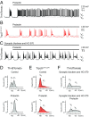
Trpc5 is required for prolactin-evoked excitation of Th+ ARC neurons. (A and B) Prolactin (500 nM) induces a tonic, long-lasting depolarization in Th+ ARC neurons from female Th-tdTomato mice (n = 10), an effect that is absent in Trpc5L3F1/L3F1 mice (n = 5). (C) Loss of prolactin (Prl)-evoked tonic depolarization (Prl, 500 nM) in a Th+ ARC neuron (Th-tdTomato) after bath application of HC-070 (100 nM) (n = 6 cells, 6 Th-tdTomato mice). Experiments were performed in the presence of a synaptic blocker mixture. IH was 0 pA in all recordings. (D) Plot of frequency distribution (recording period, 10 min) of the cell shown in A before (control) and during prolactin treatment. Control: VD = −62.4 ± 0.1 mV; VU = −53.9 ± 0.1 mV; Prl: VU = −52.1 ± 0.04 mV (mean ± SD). (E) Plot of frequency distribution (recording period, 10 min) of the cell shown in B before (control) and during prolactin treatment. Control: VD = −60.5 ± 0.03 mV; VU = −50.1 ± 0.7 mV; Prl: VD = −59.4 ± 0.1 mV; VU = −51.9 ± 0.8 mV (mean ± SD). (F) Plot of frequency distribution (recording period, 10 min) of the cell shown in C before (synaptic blockers and HC-070) and during prolactin treatment. Synaptic blockers and HC-070: VD = −60.6 ± 0.1 mV; VU = −39.9 ± 0.7 mV; Prl: VD = −56.3 ± 0.1 mV; VU = −46.1 ± 0.4 mV (mean ± SD).
Fig. 4.
Fig. 4.
Hypoprolactinemia and impaired reproductive cyclicity in Trpc5-deficient mice. (A) Examples of reproductive cycles from 7- to 12-wk-old sexually naive female Trpc5+/+ and Trpc5L3F1/L3F1 mice evaluated by daily vaginal cytology. Trpc5L3F1/L3F1 females display irregular reproductive cycles with longer dwell times in diestrus. (B) Prolonged cycle length in sexually naive Trpc5-deficient females. Trpc5+/+: 5.4 ± 0.3 d, n = 14 mice; Trpc5L3F1/L3F1: 6.5 ± 0.3 d, n = 28 mice; Trpc5-E4−/−: 8.5 ± 1.0 d, n = 5 mice; Trpc5-E5−/−: 9.7 ± 1.0 d, n = 18 mice; Kruskal–Wallis ANOVA: P < 0.001; Mann–Whitney U test: ***P < 0.001, **P < 0.01. Box plots display the interquartile ranges, median (line), and mean (black/white squares) values with whiskers indicating SD values. Each dot represents a given estrous cycle. (C and D) Checkerboard plots showing daily evaluations of vaginal smears in individual Trpc5+/+ (C) and Trpc5L3F1/L3F1 (D) females for the occurrence of estrus (filled squares). Trpc5-deficient females showed a reduced incidence of estrus. (E) Trpc5-deficient female mice harboring a global or conditional Trpc5 deletion exhibit strongly reduced Prl levels. Number of independent measurements from at least 5 mice per genotype is indicated above each box plot (Kruskal–Wallis ANOVA: P < 0.001; Mann–Whitney U test: ***P < 0.001; **P < 0.01; *P < 0.05; ns, P = 0.08 − 0.61). See table on Right for mean ± SEM values and total number of mice in parentheses. (FH) Daily analyses of prolactin levels during reproductive cycles of individual Trpc5+/+ (F) and Trpc5L3F1/L3F1 (G) mice and the group data (H) show that Trpc5L3F1/L3F1 females display prolactin surges but that overall prolactin levels are reduced. Prolactin levels are significantly diminished in Trpc5L3F1/L3F1 females at all stages of the reproductive cycle (Trpc5+/+: M, Prl, 24.7 ± 5.0 ng/mL; D, Prl, 13.9 ± 2.2 ng/mL; P, Prl, 30.2 ± 5.0 ng/mL; E, Prl, 20.4 ± 3.7 ng/mL; Trpc5L3F1/L3F1: M, Prl, 4.1 ± 1.3 ng/mL; D, Prl, 8.2 ± 2.0 ng/mL; P, Prl, 15.2 ± 3.8 ng/mL; E, Prl, 11.1 ± 3.4 ng/mL; Kruskal–Wallis ANOVA: P < 0.001; Mann–Whitney U test: ***P < 0.001). Number of independent measurements from at least 5 mice is indicated above each bar. (I) Generation and validation of Th-∆Trpc5 conditional knockout mice. Mice carrying the Trpc5-E4fx allele (Top), in which exon 4 (E4) is flanked by loxP sites (gray triangles, L1–L2), were mated with Th-Cre transgenic mice. The resulting mice carry a retained loxP site between exons 3 and 5 after deletion of exon 4. Mice were verified by PCR using genomic DNA prepared from tail (T) or hypothalamus (H), followed by sequencing of PCR products. Gel electrophoresis illustrates that genomic PCR amplifies the truncated region (232 bp) only from hypothalamus but not from tail of homozygous females (−/−) and hemizygous (–/0) males. Positive control: tail genomic DNA from Trpc4-E4−/− mice; negative controls: H2O (water template) and C57BL6/N genomic DNA (C57). PCR product sizes in base pairs are as indicated at the left. Sequencing confirmed the deletion of the exon 4 region in the hypothalamus of Th-∆Trpc5 mice. (J) Checkerboard plots showing reproductive cycles of Th-∆Trpc5 females (n = 7) reveal irregular, less frequent, and delayed estrus episodes in conditional Trpc5 knockout mice.
Fig. 5.
Fig. 5.
Impaired reproductive capabilities in Trpc5-deficient mice. (A) Relative fecundity values are strikingly reduced in Trpc5-deficient mice versus Trpc5+/+ breedings (Trpc5+/+, 17.2; Trpc5L3F1/L3F1, 2.2; Trpc5-E4−/−, 2.8; Trpc5-E5−/−, 3.9). Sexually mature (8–12 wk old) female and male mice were kept as single mating pairs over a 3-mo test period. Relative fecundity = (productive matings) × (litter size) × (number of litters). (B) Trpc5-deficient mating pairs can be fertile, but breeding success (percentage of productive matings) is diminished. Productive matings: Trpc5+/+, 16/16; Trpc5L3F1/L3F1, 13/23; Trpc5-E4−/−, 5/7; Trpc5-E5−/−, 6/8; **P < 0.01; *P < 0.05. (CE) Number of litters (C) and litter size (D) are reduced whereas the interval of pregnancies (E) increased. (C) Number of litters, Trpc5+/+: 2.3 ± 0.2; Trpc5L3F1/L3F1: 1.0 ± 0.2; Trpc5-E4−/−: 1.1 ± 0.3; Trpc5-E5−/−: 1.3 ± 0.4; Kruskal–Wallis ANOVA: P < 0.01. (D) Litter size, Trpc5+/+: 7.7 ± 0.56; Trpc5L3F1/L3F1: 3.9 ± 0.9; Trpc5-E4−/−: 3.4 ± 1.3; Trpc5-E5−/−: 4.2 ± 1.3; Kruskal-Wallis ANOVA: P < 0.05. (E) Litter interval, Trpc5+/+: 34.3 ± 3.8 d; Trpc5L3F1/L3F1: 56.8 ± 3.9 d; Trpc5-E4−/−: 50.0 ± 7.1 d; Trpc5-E5−/−: 51.0 ± 7.3 d; Kruskal–Wallis ANOVA: P < 0.01. Mann–Whitney U test: *P < 0.05; **P < 0.01; ***P < 0.001. Litter size was determined at postnatal day 0–1. Numbers in parentheses indicate number of female mice.

References

    1. Clapham D. E., Montell C., Schultz G., Julius D.; International Union of Pharmacology , International union of pharmacology. XLIII. Compendium of voltage-gated ion channels: Transient receptor potential channels. Pharmacol. Rev. 55, 591–596 (2003). - PubMed
    1. Venkatachalam K., Montell C., TRP channels. Annu. Rev. Biochem. 76, 387–417 (2007). - PMC - PubMed
    1. Zholos A., “TRPC5” in Mammalian Transient Receptor Potential (TRP) Cation Channels, Nilius B., Flockerzi V., Eds. (Springer, Berlin, Heidelberg, 2014), vol. 1, pp. 129–156.
    1. Storch U., et al. , Dynamic NHERF interaction with TRPC4/5 proteins is required for channel gating by diacylglycerol. Proc. Natl. Acad. Sci. U.S.A. 114, E37–E46 (2017). - PMC - PubMed
    1. Zhou Y., et al. , A small-molecule inhibitor of TRPC5 ion channels suppresses progressive kidney disease in animal models. Science 358, 1332–1336 (2017). - PMC - PubMed

Publication types

MeSH terms

Supplementary concepts

LinkOut - more resources