

Large distances separate coregulated genes in living Drosophila embryos
- PMID: 31285341
- PMCID: PMC6660726
- DOI: 10.1073/pnas.1908962116
Large distances separate coregulated genes in living Drosophila embryos
Abstract
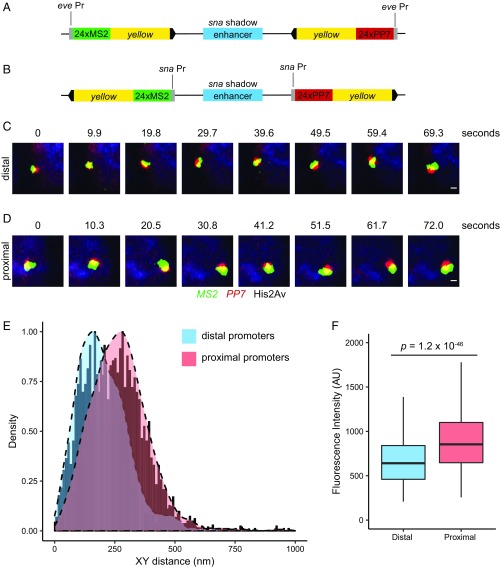
Transcriptional enhancers are short segments of DNA that switch genes on and off in response to a variety of cellular signals. Many enhancers map quite far from their target genes, on the order of tens or even hundreds of kilobases. There is extensive evidence that remote enhancers are brought into proximity with their target promoters via long-range looping interactions. However, the exact physical distances of these enhancer-promoter interactions remain uncertain. Here, we employ high-resolution imaging of living Drosophila embryos to visualize the distances separating linked genes that are coregulated by a shared enhancer. Cotransvection assays (linked genes on separate homologs) suggest a surprisingly large distance during transcriptional activity: at least 100-200 nm. Similar distances were observed when a shared enhancer was placed into close proximity with linked reporter genes in cis. These observations are consistent with the occurrence of "transcription hubs," whereby clusters (or condensates) of multiple RNA polymerase II complexes and associated cofactors are periodically recruited to active promoters. The dynamics of this process might be responsible for rapid fluctuations in the distances separating the transcription of coregulated reporter genes during transvection. We propose that enhancer-promoter communication depends on a combination of classical looping and linking models.
Keywords: Drosophila embryos; enhancers; live-imaging; transcription; transvection.
Conflict of interest statement
The authors declare no conflict of interest.
Figures


Similar articles
-
Visualization of Transvection in Living Drosophila Embryos.Mol Cell. 2018 Apr 19;70(2):287-296.e6. doi: 10.1016/j.molcel.2018.02.029. Epub 2018 Mar 29. Mol Cell. 2018. PMID: 29606591 Free PMC article.
-
Enhancer Control of Transcriptional Bursting.Cell. 2016 Jul 14;166(2):358-368. doi: 10.1016/j.cell.2016.05.025. Epub 2016 Jun 9. Cell. 2016. PMID: 27293191 Free PMC article.
-
Temporal dissection of an enhancer cluster reveals distinct temporal and functional contributions of individual elements.Mol Cell. 2021 Mar 4;81(5):969-982.e13. doi: 10.1016/j.molcel.2020.12.047. Epub 2021 Jan 21. Mol Cell. 2021. PMID: 33482114
-
Enhancer-promoter communication in Drosophila developmental gene transcription.Int J Dev Biol. 2024;68(4):169-188. doi: 10.1387/ijdb.230218gh. Int J Dev Biol. 2024. PMID: 38869221 Review.
-
One thousand and one ways of making functionally similar transcriptional enhancers.Bioessays. 2008 Nov;30(11-12):1052-7. doi: 10.1002/bies.20849. Bioessays. 2008. PMID: 18937349 Review.
Cited by
-
3D enhancer-promoter interactions and multi-connected hubs: Organizational principles and functional roles.Cell Rep. 2023 Apr 25;42(4):112068. doi: 10.1016/j.celrep.2023.112068. Epub 2023 Apr 13. Cell Rep. 2023. PMID: 37059094 Free PMC article. Review.
-
Independence of chromatin conformation and gene regulation during Drosophila dorsoventral patterning.Nat Genet. 2021 Apr;53(4):487-499. doi: 10.1038/s41588-021-00799-x. Epub 2021 Apr 1. Nat Genet. 2021. PMID: 33795866 Free PMC article.
-
New insights into promoter-enhancer communication mechanisms revealed by dynamic single-molecule imaging.Biochem Soc Trans. 2021 Jun 30;49(3):1299-1309. doi: 10.1042/BST20200963. Biochem Soc Trans. 2021. PMID: 34060610 Free PMC article. Review.
-
The Role of Insulation in Patterning Gene Expression.Genes (Basel). 2019 Sep 28;10(10):767. doi: 10.3390/genes10100767. Genes (Basel). 2019. PMID: 31569427 Free PMC article. Review.
-
RNA polymerase II transcription compartments - from factories to condensates.Nat Rev Genet. 2025 Jun 19. doi: 10.1038/s41576-025-00859-6. Online ahead of print. Nat Rev Genet. 2025. PMID: 40537661 Review.
References
-
- Cisse I. I., et al. , Real-time dynamics of RNA polymerase II clustering in live human cells. Science 341, 664–667 (2013). - PubMed
Publication types
MeSH terms
Substances
Grants and funding
LinkOut - more resources
Full Text Sources
Molecular Biology Databases
