. 2019 Jul 8;9(1):9850.
doi: 10.1038/s41598-019-46367-6.

Blockade of L-type Ca2+ channel attenuates doxorubicin-induced cardiomyopathy via suppression of CaMKII-NF-κB pathway

Affiliations

Blockade of L-type Ca2+ channel attenuates doxorubicin-induced cardiomyopathy via suppression of CaMKII-NF-κB pathway

Soichiro Ikeda et al. Sci Rep. .

Abstract

Ca2+/calmodulin-dependent protein kinase II (CaMKII) and nuclear factor-kappa B (NF-κB) play crucial roles in pathogenesis of doxorubicin (DOX)-induced cardiomyopathy. Their activities are regulated by intracellular Ca2+. We hypothesized that blockade of L-type Ca2+ channel (LTCC) could attenuate DOX-induced cardiomyopathy by regulating CaMKII and NF-κB. DOX activated CaMKII and NF-κB through their phosphorylation and increased cleaved caspase 3 in cardiomyocytes. Pharmacological blockade or gene knockdown of LTCC by nifedipine or small interfering RNA, respectively, suppressed DOX-induced phosphorylation of CaMKII and NF-κB and apoptosis in cardiomyocytes, accompanied by decreasing intracellular Ca2+ concentration. Autocamtide 2-related inhibitory peptide (AIP), a selective CaMKII inhibitor, inhibited DOX-induced phosphorylation of NF-κB and cardiomyocyte apoptosis. Inhibition of NF-κB activity by ammonium pyrrolidinedithiocarbamate (PDTC) suppressed DOX-induced cardiomyocyte apoptosis. DOX-treatment (18 mg/kg via intravenous 3 injections over 1 week) increased phosphorylation of CaMKII and NF-κB in mouse hearts. Nifedipine (10 mg/kg/day) significantly suppressed DOX-induced phosphorylation of CaMKII and NF-κB and cardiomyocyte injury and apoptosis in mouse hearts. Moreover, it attenuated DOX-induced left ventricular dysfunction and dilatation. Our findings suggest that blockade of LTCC attenuates DOX-induced cardiomyocyte apoptosis via suppressing intracellular Ca2+ elevation and activation of CaMKII-NF-κB pathway. LTCC blockers might be potential therapeutic agents against DOX-induced cardiomyopathy.


Conflict of interest statement

The authors declare no competing interests.

Figures

Figure 1
Figure 1
DOX induced phosphorylation of CaMKII and NF-κB and increased cleaved caspase 3 in cardiomyocytes in a dose-dependent manner. (a) Representative immunoblots of CaMKII, phosphorylated CaMKII, NF-κB, phosphorylated NF-κB, cleaved caspase 3, and GAPDH in cultured neonatal rat ventricular myocytes (NRVMs) treated with indicated concentrations of DOX. (bd) Quantitative analysis of phosphorylated CaMKII, phosphorylated NF-κB, and cleaved caspase 3 in NRVMs treated with indicated concentration of DOX (n = 5). The experiment was conducted 2 times. *P < 0.05: post-hoc Tukey’s comparison test.
Figure 2
Figure 2
Blockade of LTCC suppressed DOX-induced phosphorylation of CaMKII and NF-κB and increases in cleaved caspase 3 in cardiomyocytes. (a) Representative immunoblots of CaMKII, phosphorylated CaMKII, NF-κB, phosphorylated NF-κB, cleaved caspase 3, and GAPDH in NRVMs treated with or without nifedipine (Nif, 1 μM) in the presence or absence of DOX (10 μM) for 24 hour (n = 5). The experiment was conducted 3 times. (bd) Quantitative analysis of phosphorylated CaMKII, phosphorylated NF-κB, and cleaved caspase 3 in each group (n = 5). (e) Representative immunoblots of CaMKII, phosphorylated CaMKII, NF-κB, phosphorylated NF-κB, cleaved caspase 3, and GAPDH in NRVMs treated with indicated small interfering RNA (siRNA) in the presence or absence of DOX (10 μM) for 24 hour (n = 5). (fh) Quantitative analysis of phosphorylated CaMKII, phosphorylated NF-κB, and cleaved caspase 3 in each group (n = 5). The experiment was conducted 3 times. *P < 0.05, **P < 0.01: post-hoc Tukey’s comparison test.
Figure 3
Figure 3
Blockade of LTCC attenuated DOX-induced cardiomyocyte apoptosis. (a,b) Apoptosis evaluated by TUNEL staining in NRVMs treated with nifedipine (Nif, 1 μM) or siRNA for LTCC (1 nM) in the presence or absence of DOX (10 μM) for 24 hours (n = 5–6). The experiment was conducted 3 times. Arrows indicate TUNEL-positive cells. (c) LDH release, a marker of cell death, evaluated by LDH cytotoxicity assay in NRVMs treated with nifedipine (Nif) or siRNA for LTCC in the presence or absence of DOX (10 μM) for 24 hours (n = 5–6). The experiment was conducted 3 times. **P < 0.01: post-hoc Tukey’s comparison test.
Figure 4
Figure 4
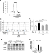
Blockade of LTCC attenuated DOX-induced elevation of intracellular Ca2+ levels in cardiomyocytes. (a) The resting levels of intracellular Ca2+ concentration in NRVMs were evaluated by fura-2 fluorometry. (b) A representative recording of fura-2 fluorometry in NRVMs treated with nifedipine (Nif) or siRNA for LTCC in the presence or absence of DOX (10 μM) for 24 hours. (c) The resting levels of intracellular Ca2+ were expressed as percentages by assigning the levels of intracellular Ca2+ obtained with ionomycin in the presence and absence of extracellular Ca2+, to be 100% and 0%, respectively. Cap-tipped lines indicate intracellular Ca2+ levels (n = 5–8). (d) Representative immunoblots of CaMKII, phosphorylated CaMKII, and GAPDH in NRVMs treated with or without BAPTA (2 μM) in the presence or absence of DOX (10 μM) for 24 hour (n = 5). (e) Quantitative analysis of phosphorylated CaMKII in each group (n = 5). The experiment was conducted 2 times. **P < 0.01: post-hoc Tukey’s comparison test.
Figure 5
Figure 5
CaMKII positively regulated DOX-induced cardiomyocyte apoptosis by activating NF-κB. (a) Representative immunoblots of CaMKII, phosphorylated CaMKII, NF-κB, phosphorylated NF-κB, cleaved caspase 3, and GAPDH in NRVMs treated with or without AIP (10 μM, 24 h) in the presence or absence of DOX (10 μM) for 24 hours (n = 5–6). (bd) Quantitative analysis of phosphorylated CaMKII, phosphorylated NF-κB, and cleaved caspase 3 in each group (n = 5–6). The experiment was conducted 3 times. (e) Apoptosis evaluated by TUNEL staining in each group (n = 5). The experiment was conducted 2 times. (f) Representative immunoblots of NF-κB, phosphorylated NF-κB, cleaved caspase 3, and GAPDH in NRVMs treated with or without PDTC (100 μM, 24 h) in the presence or absence of DOX (10 μM) for 24 hours (n = 5–6). (gi) Quantitative analysis of phosphorylated NF-κB and cleaved caspase 3 in NRVMs treated with or without PDTC (100 μM, 24 h) in the presence or absence of DOX (10 μM) for 24 hours (n = 5–6). The experiment was conducted 2 times. (j) Apoptosis evaluated by TUNEL staining in each group (n = 5). The experiment was conducted 2 times. *P < 0.05, **P < 0.01: post-hoc Tukey’s comparison test.
Figure 6
Figure 6
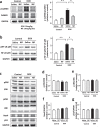
Blockade of LTCC suppressed CaMKII-NF-kB pathway in DOX-treated hearts. (a) Representative immunoblots and quantitative analysis of CaMKII, phosphorylated CaMKII, and GAPDH in DOX (3 doses of DOX at 6 mg/kg body weight every third day for 1 week) or control vehicle (phosphate-buffered saline: PBS) treated-C57B/6 J mouse hearts subjected to either nifedipine (Nif, 10 mg/day/day) or saline for 9 days (n = 5). (b) Representative immunoblots and quantitative analysis of NF-κB, phosphorylated NF-κB, cleaved caspase 3, and GAPDH in each group (n = 5). The experiment was conducted 3 times. (cg) Representative immunoblotsa and quantitative analysis of ERK, phosphorylated ERK, JNK, phosphorylated JNK, Nox4, p53, and GAPDH in each group (n = 5).
Figure 7
Figure 7
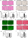
Blockade of LTCC ameliorated DOX-induced myocardial injury and apoptosis in mice. (a) HE stained section in DOX (3 doses of DOX at 6 mg/kg body weight every third day for 1 week) or control vehicle (phosphate-buffered saline: PBS) treated-C57B/6 J mouse hearts subjected to either nifedipine (Nif, 10 mg/day/day) or saline for 14 days. Arrows indicate cytoplasmic vacuolization and arrowheads indicate myofibrillar loss. (b) Cardiomyocyte injury as assessed by number of cytoplasmic vaculolization (n = 6). (c) Cardiomyocyte death as assessed by Billingham score in the heart (n = 5). (d) TUNEL stained heart section in each group. Arrow heads indicate TUNEL-positive nuclei. (e) The percentage of TUNEL-positive nuclei in the heart (n = 5). (f) WGA-stained heart section in each group. (g) Cardiomyocyte hypertrophy as assessed by cross-sectional area in each group (n = 5). (h) Masson-trichrome-stained heart section in each group. (i) Interstitial fibrosis as assessed by collagen volume in each group (n = 5). *P < 0.05, **P < 0.01: post-hoc Tukey’s comparison test.
Figure 8
Figure 8
Blockade of LTCC attenuated DOX-induced cardiac dysfunction and loss of heart weight in mice. (a) The representative echocardiographic images of DOX (3 doses of DOX at 6 mg/kg body weight every third day for 1 week) or control vehicle (phosphate-buffered saline: PBS) treated-C57B/6 J mouse hearts subjected to either nifedipine (Nif, 10 mg/day/day) or saline for 14 days. Long two-way arrows and short two-way arrows indicate left ventricular end-diastolic diameter (LVDd) and left ventricular end-systolic diameter (LVDs), respectively. (bf) Heart rate, LVDd and LVDs, fractional shortening, and LV ejection fraction measured by echocardiography at day14 in each group (n = 6–14). (g,h) Heart weight to tibial length (TL) ratio and LV weight to TL ratio in each group (n = 6–14). *P < 0.05, **P < 0.01: post-hoc Tukey’s comparison test.

Similar articles

Cited by

References

    1. Barrett-Lee PJ, et al. Expert opinion on the use of anthracyclines in patients with advanced breast cancer at cardiac risk. Ann Oncol. 2009;20:816–827. doi: 10.1093/annonc/mdn728. - DOI - PubMed
    1. Felker GM, et al. Underlying causes and long-term survival in patients with initially unexplained cardiomyopathy. N Engl J Med. 2000;342:1077–1084. doi: 10.1056/NEJM200004133421502. - DOI - PubMed
    1. Kotamraju S, Konorev EA, Joseph J, Kalyanaraman B. Doxorubicin-induced apoptosis in endothelial cells and cardiomyocytes is ameliorated by nitrone spin traps and ebselen. Role of reactive oxygen and nitrogen species. J Biol Chem. 2000;275:33585–33592. doi: 10.1074/jbc.M003890200. - DOI - PubMed
    1. Wang L, Ma W, Markovich R, Chen JW, Wang PH. Regulation of cardiomyocyte apoptotic signaling by insulin-like growth factor I. Circ Res. 1998;83:516–522. doi: 10.1161/01.RES.83.5.516. - DOI - PubMed
    1. Zhang S, et al. Identification of the molecular basis of doxorubicin-induced cardiotoxicity. Nat Med. 2012;18:1639–1642. doi: 10.1038/nm.2919. - DOI - PubMed

Publication types

MeSH terms