. 2019 Aug;26(8):704-711.
doi: 10.1038/s41594-019-0262-6. Epub 2019 Jul 8.

Structure of the eukaryotic protein O-mannosyltransferase Pmt1-Pmt2 complex

Affiliations

Structure of the eukaryotic protein O-mannosyltransferase Pmt1-Pmt2 complex

Lin Bai et al. Nat Struct Mol Biol. 2019 Aug.

Abstract

In eukaryotes, a nascent peptide entering the endoplasmic reticulum (ER) is scanned by two Sec61 translocon-associated large membrane machines for protein N-glycosylation and protein O-mannosylation, respectively. While the structure of the eight-protein oligosaccharyltransferase complex has been determined recently, the structures of mannosyltransferases of the PMT family, which are an integral part of ER protein homeostasis, are still unknown. Here we report cryo-EM structures of the Saccharomyces cerevisiae Pmt1-Pmt2 complex bound to a donor and an acceptor peptide at 3.2-Å resolution, showing that each subunit contains 11 transmembrane helices and a lumenal β-trefoil fold termed the MIR domain. The structures reveal the substrate recognition model and confirm an inverting mannosyl-transferring reaction mechanism by the enzyme complex. Furthermore, we found that the transmembrane domains of Pmt1 and Pmt2 share a structural fold with the catalytic subunits of oligosaccharyltransferases, confirming a previously proposed evolutionary relationship between protein O-mannosylation and protein N-glycosylation.


Conflict of interest statement

Competing Interests

The authors declare no competing financial interests.

Figures
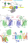
Fig. 1.
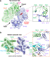
Fig. 1.. Structure of the yeast Pmt1–Pmt2 complex.
(a) Both protein O-mannosylation and protein N-glycosylation can be carried out co-translationally and co-translocationally, because both Pmt1–Pmt2 and OST physically associate with the Sec61 translocon. (b) The overall structure of Pmt1–Pmt2 in cartoons is shown in front and back views (green for Pmt1 and purple-blue for Pmt2). The Dol-P and N-glycans are shown as orange and red sticks, respectively. The acceptor peptide is shown as red spheres. (c) A ribbon diagram and topological sketch of Pmt1. The 11 TMHs are numbered and colored in a rainbow scheme. The MIR domain is drawn as a green circle. The number and identity of the first residue on each TMH is labeled. The red asterisk marks the DE motif at the beginning of the first horizontal helix (HH1).
Fig. 2.
Fig. 2.. The interfaces between Pmt1 and Pmt2.
A lumenal view (a) and a cytosolic view (b) of the Pmt1–Pmt2 structure. The interface areas between the two subunits are highlighted by a dashed black rectangle in (a) and by a dashed orange rectangle in (b). The areas in the rectangles are enlarged at the right. The lipid molecule mediating the Pmt1–Pmt2 interaction at the cytosolic side is shown as sticks.
Fig. 3.
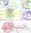
Fig. 3.. The active site of the Pmt1–Pmt2 complex.
(a) Pmt1 is shown as a cartoon and a transparent surface view. The transmembrane region of Pmt2 (purple-blue) is superposed with Pmt1. Dol-P is shown as orange sticks, and the acceptor peptide is shown as a red cartoon. (b) Close-up view of the highly conserved DE motif of Pmt1 and Pmt2. (c) Close-up view of the active site of Pmt1. (d) Close-up view of the active site of Pmt2. (e) LIGPLOT scheme for Dol-P and acceptor peptide binding residues of Pmt1. The pink circle represents the active site of the enzyme.
Fig. 4.
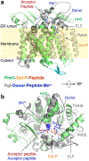
Fig. 4.. Superimposition of the transmembrane domain of Pmt1 (green) with that of the PglB structure (grey).
A side view (a) and a top view (b) of the superimposition of the transmembrane domain of Pmt1 (green) with that of the PglB structure (PDB ID 5OGL, grey), which is in complex with an acceptor peptide, Mn2+, and a nonhydrolyzable lipid-linked oligosaccharide analog, (ωZZZ)-PPC-GlcNAc (blue). The red asterisk denotes the overlapping phosphate groups and the acceptor residues in the two structures. The acceptor residues, a Thr in Pmt1–Pmt2 and an Asn in PglB, are located on the opposite side of the donor group, in consistent with the fact that both enzymes are inverting glycosyltransferases.
Fig. 5.
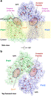
Fig. 5.. Mapping of the congenital muscular dystrophy mutations found in human POMT1–POMT2 on to the structure of the yeast Pmt1–Pmt2.
(a, b) A side and a top lumenal views of the Pmt1-Pmt2 in ribbon presentation. The Cα atoms of disease-related residues are shown as salmon spheres. The active sites are shown by transparent pink ovals. The acceptor peptide and Dol-P are shown as sticks. Individual mutations are summarized in Supplementary Table 1.

Similar articles

Cited by

References

    1. Panin VM & Wells L Protein O-mannosylation in metazoan organisms. Curr Protoc Protein Sci 75, 12–12 (2014). - PMC - PubMed
    1. VanderVen BC, Harder JD, Crick DC & Belisle JT Export-mediated assembly of mycobacterial glycoproteins parallels eukaryotic pathways. Science 309, 941–3 (2005). - PubMed
    1. Xu C, Wang S, Thibault G & Ng DT Futile protein folding cycles in the ER are terminated by the unfolded protein O-mannosylation pathway. Science 340, 978–81 (2013). - PubMed
    1. Loibl M & Strahl S Protein O-mannosylation: what we have learned from baker’s yeast. Biochim Biophys Acta 1833, 2438–46 (2013). - PubMed
    1. Burda P & Aebi M The dolichol pathway of N-linked glycosylation. Biochim Biophys Acta 1426, 239–57 (1999). - PubMed

Methods-only References

    1. Otwinowski Z & Minor W Processing of X-ray diffraction data collected in oscillation mode. Methods Enzymol 276, 307–326 (1997). - PubMed
    1. Adams PD et al. PHENIX: a comprehensive Python-based system for macromolecular structure solution. Acta Crystallogr D Biol Crystallogr 66, 213–221 (2010). - PMC - PubMed
    1. Emsley P, Lohkamp B, Scott WG & Cowtan K Features and development of Coot. Acta Crystallogr D Biol Crystallogr 66, 486–501 (2010). - PMC - PubMed
    1. Murshudov GN, Vagin AA & Dodson EJ Refinement of macromolecular structures by the maximum-likelihood method. Acta Crystallogr D Biol Crystallogr 53, 240–255, doi:10.1107/S0907444996012255 (1997). - DOI - PubMed
    1. Zheng SQ et al. MotionCor2: anisotropic correction of beam-induced motion for improved cryo-electron microscopy. Nat Methods 14, 331–332 (2017). - PMC - PubMed

Publication types

MeSH terms