. 2019 Aug;26(8):679-685.
doi: 10.1038/s41594-019-0258-2. Epub 2019 Jul 8.

Cryo-EM structures reveal coordinated domain motions that govern DNA cleavage by Cas9

Affiliations

Cryo-EM structures reveal coordinated domain motions that govern DNA cleavage by Cas9

Xing Zhu et al. Nat Struct Mol Biol. 2019 Aug.

Abstract

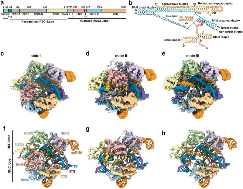
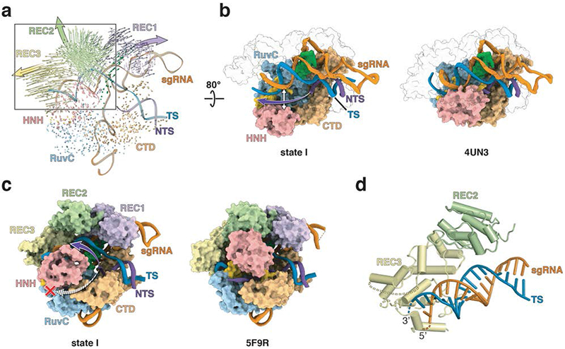
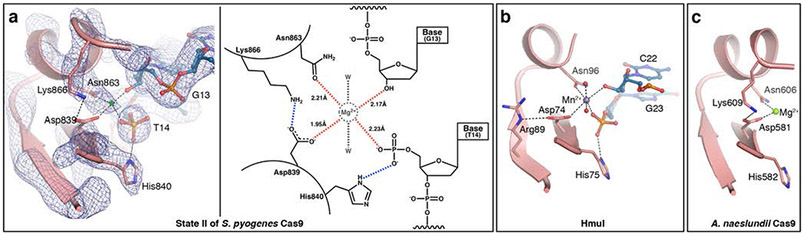
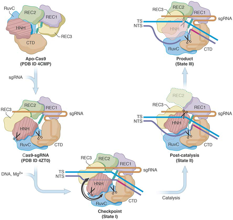
The RNA-guided Cas9 endonuclease from Streptococcus pyogenes is a single-turnover enzyme that displays a stable product state after double-stranded-DNA cleavage. Here, we present cryo-EM structures of precatalytic, postcatalytic and product states of the active Cas9-sgRNA-DNA complex in the presence of Mg2+. In the precatalytic state, Cas9 adopts the 'checkpoint' conformation with the HNH nuclease domain positioned far away from the DNA. Transition to the postcatalytic state involves a dramatic ~34-Å swing of the HNH domain and disorder of the REC2 recognition domain. The postcatalytic state captures the cleaved substrate bound to the catalytically competent HNH active site. In the product state, the HNH domain is disordered, REC2 returns to the precatalytic conformation, and additional interactions of REC3 and RuvC with nucleic acids are formed. The coupled domain motions and interactions between the enzyme and the RNA-DNA hybrid provide new insights into the mechanism of genome editing by Cas9.


Figures

Fig. 1.
Fig. 1.. Cryo-EM structures of three states of Cas9-sgRNA-dsDNA complex.
(a) Domain organization of S. pyogenes Cas9. (b) Schematic diagram of the nucleic acids used in the study. The sgRNA is orange, and the target (TS) and non-target strands (NTS) of the dsDNA are blue and purple, respectively. Cryo-EM density maps (c-e) and schematic representations (f-h) of states I, II and III of the Cas9-sgRNA-dsDNA ternary complex. Protein domains and nucleic acids are colored as in (a) and (b). Some regions of weak density, shown in grey in panels c-e, are not included in the atomic models shown in panels f-h.
Fig. 2.
Fig. 2.. The central channel of Cas9 accommodates the R-loop structure in state I.
(a) Superposition of Cas9-sgRNA (PDB ID 4ZT0) onto state I, with RuvC as the common reference, reveals the extent of Cas9 domain movements upon dsDNA and Mg2+ binding. Arrow lengths correspond to the magnitude of Cα atom movements; arrowheads show domain movement directions and large solid arrows point to the general direction of the domain movement. (b) The central channel in state I (left panel) and Cas9 bound to the partial duplex (right panel; PDB ID 4UN3). The purple arrow (left panel) indicates the trajectory for the disordered NTS. The helical recognition lobes are shown as transparent surfaces for clarity. (c) The central channel in state I (left panel) and the “Mg2+-free” complex (right panel; PDB ID 5F9R). The purple and striped arrows (left panel) indicate two possible trajectories for the disordered NTS. The red cross indicates that the tunnel between HNH and RuvC is closed in state I. (d) A global view showing interactions between REC3 domain and the sgRNA-TS duplex in the PAM-distal. Dashed lines indicate unmodeled regions of L1 and REC3. The domains and nucleic acids are colored as in Figs. 1a, b.
Fig. 3.
Fig. 3.. HNH domain adopts catalytic conformation in state II.
(a) A conformational change of the HNH domain during transition from state I to state II. The HNH domain in state I (beige cartoon) rotates around a central axis (grey rod) and translates ~34 Å to reach the scissile bond in TS (shown with dashed circle, and either arrow or asterisk) where it adopts an active conformation (pink cartoon). The nucleic acid backbone is colored as in Fig. 1 with the rest of Cas9 shown in grey transparent surface. (b) Close-up view of the catalytically-competent active site of HNH in state II with ball-and-stick representation of the catalytic site. The newly formed 5’- and 3’-ends of the cleaved TS are shown in blue, with hydrogen bonds shown as dashed lines. (c) Cryo-EM density maps and models spanning the cleavage site demonstrate that the TS is intact in state I and cleaved in states II and III. Arrow designates the scissile bond in state I, while the asterisk marks the cleaved phosphodiester bond in states II and III.
Fig. 4.
Fig. 4.. The HNH active site conformation in state II.
(a) Left: Close-up view of the active site of the HNH domain in state II (pink) with the corresponding cryo-EM map (blue mesh) superposed. A putative metal ion position is marked with green asterisk. Right: Schematic representation of the state II HNH active site. Putative Mg2+ ion, water molecules and their interactions are colored grey. Observed H-bonds are blue dashed lines, while red dashed lines designate putative interactions between Cas9 side chains and unmodeled Mg2+. Distances between coordinating Cas9 residues and the proposed Mg2+ion location are shown in Å. (b) The active site of the homing endonuclease I, HmuI (PDB ID 1U3E) . (c) The active site of the HNH domain from A. naeslundii Cas9 (PDB ID 4OGE) . In (a, left), (b) and (c), catalytic residues and reaction products are shown as pink and blue sticks, respectively, and hydrogen bonds, both observed and putative, are shown as dashed lines. In panels b and c, purple, green and red spheres are Mn2+, Mg2+ and water molecules, respectively.
Fig. 5.
Fig. 5.. Proposed mechanism for the concerted series of domain movements involved in Cas9-mediated DNA cleavage.
The binding of sgRNA to apo-Cas9 induces major domain rearrangements and formation of the binary complex. In the presence of dsDNA and Mg2+, the “checkpoint” conformation (State I) is formed. Target strand base-pairs with NTS and runs parallel to sgRNA, HNH adopts an inactive conformation, and the distal DNA duplex is short. Upon activation, HNH rotates and swings (arrows indicate possible routes) towards the cleavage site in TS. This is accompanied by REC2 disorder, ordering of REC3 loops that bind to the distal PAM, and interactions between RuvC and the longer distal DNA duplex. During catalysis, the HNH and RuvC active sites (scissors) cleave the TS and NTS, respectively. After cleavage, HNH remains bound to products, whereas the RuvC active site is near the cleaved NTS. This arrangement is captured in the “post-catalytic” complex (State II). In the “product” complex (State III), HNH dissociates from the cleaved TS and becomes disordered, REC2 is ordered and adopts its State I conformation, while interactions between REC3, RuvC and nucleic acids persist. Apo-Cas9 and Cas9-sgRNA are based on PDB ID 4CMP and 4ZT0 , respectively.

References

    1. Cong L et al. Multiplex genome engineering using CRISPR/Cas systems. Science 339, 819–823, doi:10.1126/science.1231143 (2013). - DOI - PMC - PubMed
    1. Mali P et al. RNA-guided human genome engineering via Cas9. Science 339, 823–826, doi:10.1126/science.1232033 (2013). - DOI - PMC - PubMed
    1. Boyle EA et al. High-throughput biochemical profiling reveals sequence determinants of dCas9 off-target binding and unbinding. Proc Natl Acad Sci U S A 114, 5461–5466, doi:10.1073/pnas.1700557114 (2017). - DOI - PMC - PubMed
    1. Sternberg SH, LaFrance B, Kaplan M & Doudna JA Conformational control of DNA target cleavage by CRISPR-Cas9. Nature 527, 110–113, doi:10.1038/nature15544 (2015). - DOI - PMC - PubMed
    1. Dagdas YS, Chen JS, Sternberg SH, Doudna JA & Yildiz A A conformational checkpoint between DNA binding and cleavage by CRISPR-Cas9. Sci Adv 3, eaao0027, doi:10.1126/sciadv.aao0027 (2017). - DOI - PMC - PubMed

Methods-only References:

    1. Anders C & Jinek M In vitro enzymology of Cas9. Methods Enzymol 546, 1–20, doi:10.1016/B978-0-12-801185-0.00001-5 (2014). - DOI - PMC - PubMed
    1. Scheres SH RELION: implementation of a Bayesian approach to cryo-EM structure determination. J Struct Biol 180, 519–530, doi:10.1016/j.jsb.2012.09.006 (2012). - DOI - PMC - PubMed
    1. Zheng SQ et al. MotionCor2: anisotropic correction of beam-induced motion for improved cryo-electron microscopy. Nat Methods 14, 331–332, doi:10.1038/nmeth.4193 (2017). - DOI - PMC - PubMed
    1. Punjani A, Rubinstein JL, Fleet DJ & Brubaker MA cryoSPARC: algorithms for rapid unsupervised cryo-EM structure determination. Nat Methods 14, 290–296, doi:10.1038/nmeth.4169 (2017). - DOI - PubMed
    1. Adams PD et al. PHENIX: a comprehensive Python-based system for macromolecular structure solution. Acta Crystallogr D Biol Crystallogr 66, 213–221, doi:10.1107/S0907444909052925 (2010). - DOI - PMC - PubMed

Publication types