. 2019 Jun 22:10:12.
doi: 10.1186/s13227-019-0126-5. eCollection 2019.

Reinvestigating the early embryogenesis in the flatworm Maritigrella crozieri highlights the unique spiral cleavage program found in polyclad flatworms

Affiliations

Reinvestigating the early embryogenesis in the flatworm Maritigrella crozieri highlights the unique spiral cleavage program found in polyclad flatworms

Johannes Girstmair et al. Evodevo. .

Abstract

Background: Spiral cleavage is a conserved, early developmental mode found in several phyla of Lophotrochozoans resulting in highly diverse adult body plans. While the cleavage pattern has clearly been broadly conserved, it has also undergone many modifications in various taxa. The precise mechanisms of how different adaptations have altered the ancestral spiral cleavage pattern are an important ongoing evolutionary question, and adequately answering this question requires obtaining a broad developmental knowledge of different spirally cleaving taxa. In flatworms (Platyhelminthes), the spiral cleavage program has been lost or severely modified in most taxa. Polyclad flatworms, however, have retained the pattern up to the 32-cell stage. Here we study early embryogenesis of the cotylean polyclad flatworm Maritigrella crozieri to investigate how closely this species follows the canonical spiral cleavage pattern and to discover any potential deviations from it.

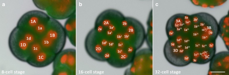
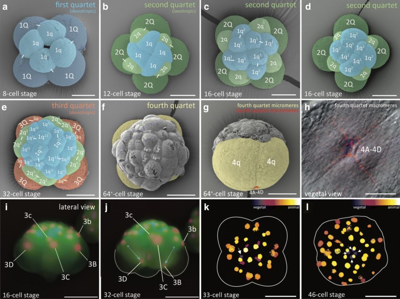
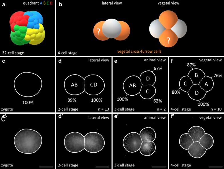
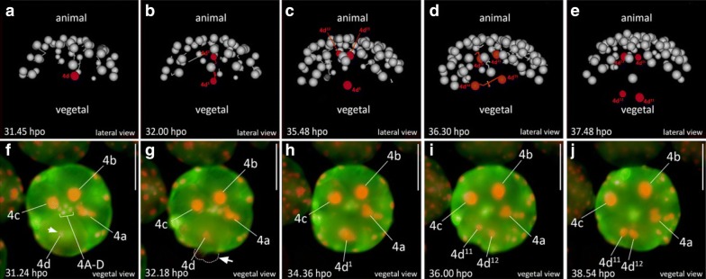
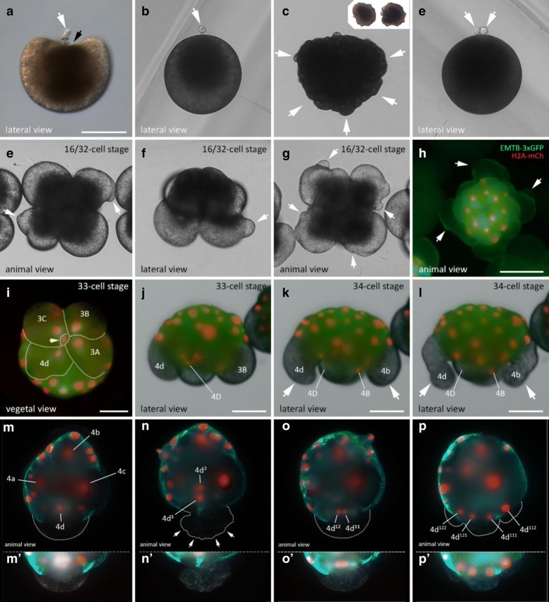
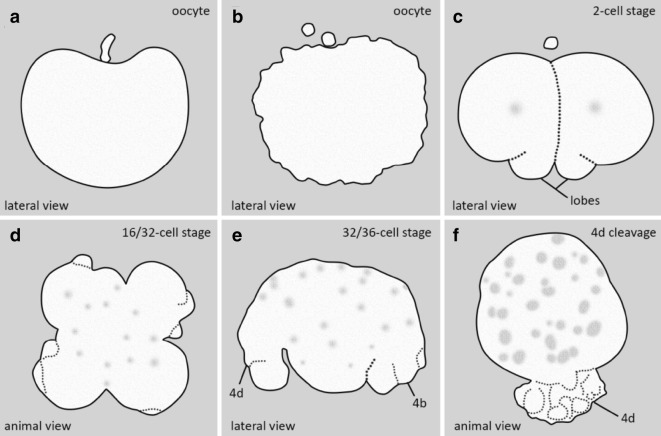
Results: Using live imaging recordings and 3D reconstructions of embryos, we give a detailed picture of the events that occur during spiral cleavage in M. crozieri. We suggest, contrary to previous observations, that the four-cell stage is a product of unequal cleavages. We show that that the formation of third and fourth micromere quartets is accompanied by strong blebbing events; blebbing also accompanies the formation of micromere 4d. We find an important deviation from the canonical pattern of cleavages with clear evidence that micromere 4d follows an atypical cleavage pattern, so far exclusively found in polyclad flatworms.

Conclusions: Our findings highlight that early development in M. crozieri deviates in several important aspects from the canonical spiral cleavage pattern. We suggest that some of our observations extend to polyclad flatworms in general as they have been described in both suborders of the Polycladida, the Cotylea and Acotylea.

Keywords: Blebbing; Evo-devo; Light-sheet microscopy; Live imaging; Polyclad flatworms; SPIM; Spiralians; Symmetry breaking; Turbellarians.


Conflict of interest statement

Competing interestsThe authors declare that they have no competing interests.

Figures

Fig. 1
Fig. 1
Schematics and nomenclature of the spiral quartet cleavage as found in polyclad flatworms. Micromere and macromere quartets (q and Q, respectively) are colour-coded. a The third cleavage (four- to eight-cell stage) is unequal and asymmetric. The eight-cell stage embryo consists of four larger vegetal macromeres 1Q, and four smaller animally positioned micromeres 1q sitting skewed to one side of their sister macromere, above the macromeres’ cleavage furrows. The typical spiral deformations (SD) of macromeres show a helical twist towards one side with respect to the animal–vegetal axis. This is best seen if the embryo is viewed from the animal pole. The resulting spiral shape taken by all four macromeres has been shown to be either clockwise (dexiotropic) or counter clockwise (laeotropic) among different lophotrochozoans. In the polyclad M. crozieri it is dexiotropic. Notably it has been demonstrated that the mechanism of spiral deformations depends on actin filaments rather than on spindle forming microtubules [70]. b, c In subsequent rounds of division, the larger macromeres again divide unequally and asymmetrically sequentially forming the second and then the third quartets of micromeres. During these divisions the spiral deformations appear in alternating dexiotropic/laeotropic directions (the rule of alternation). Up to the 32-cell stage, polyclad flatworms represent a classic example of stereotypic lophotrochozoan spiral quartet cleavage. d The formation of the fourth quartet (4Q and 4q) deviates from the typical pattern seen in other spiral-cleaving embryos insofar as the micromeres 4q become large and the macromeres 4Q diminutive. Q = A, B, C, D; q = a, b, c, d
Fig. 2
Fig. 2
Live imaging of the transition from an8-cell stage embryo to a 32-cell stage in M. crozieri with nuclei labelled according to the canonical spiral cleavage nomenclature. a The eight-cell stage is a product of a dexiotropic cleavage. b The 16-cell stage with its first quartet micromeres (after their first cleavage round) and second quartet micromeres (2a–2d) and macromeres (2A–2D). c The embryo has now reached the 32-cell stage. Images captured with a Zeiss Axio Zoom.V16 Stereo Microscope. Scale bar is 50 µm
Fig. 3
Fig. 3
Formation of the four quartets in M. crozieri. ag SEM pictures coloured according to micromere quartets. a First quartet (1Q and 1q indicated in blue). bd Second quartet (2Q and 2q) indicated in green. e Third quartet (3Q and 3q) indicated in orange. f, g The large fourth micromere quartet (4q) are shown and indicated in yellow. The fourth quartet micromeres are shown in red. h Closeup of the fourth quartet micromeres (4A–4D) with Phalloidin staining (red) outlining their cell shape. Nuclear staining (blue) is DAPI. il Formation of the fourth quartet. i The 16-cell stage shows macromeres 3B-D and their nuclei at an animal position within the large blastomeres. j Same embryo as in G but at the 32-cell stage. Nuclei of 3B and 3D are now positioned at the vegetal pole of the macromeres. k 33-cell stage of a 3D reconstructed embryo (Their depth in the embryo is coded by colours as seen in top right part of the panel. Division of one of the four macromeres (3Q) into 4Q/4q has taken place. The white arrow indicates the newly formed small macromere of the fourth quartet (4Q) coloured purple indicating it is close to the vegetal pole. l 3D reconstructions showing that all four macromeres comprising the fourth quartet are now positioned at the most vegetal pole of the embryo (coloured purple and indicated by arrows). Scale bar sare 50 μm
Fig. 4
Fig. 4
Putative cell–cell interactions observed in the gastrulating polyclad flatworm M. crozieri. ai A descendant of cell 4d1 (red arrow) is traced and can be seen approaching and later departing from fourth quartet macromeres 4A-D before epiboly is completed. Time represents hours (h) of time-lapse imaging with an OpenSPIM. Scale bars are50 µm
Fig. 5
Fig. 5
Averaged volume measurements in M. crozieri blastomeres of the first and second cleavages. a A 3D model of a 32-cell stage embryo is shown with descendants derived from the same four-cell blastomere indicated by the same colour. b A 3D model of a four-cell stage embryo is depicted showing both vegetal cross-furrow cells that meet at the vegetal pole indicated in orange. Whether the D quadrant is already specified in M. crozieri at the four-cell stage remains unclear, which is indicated here by a question mark. cf Volumes are given as a percentage of the volume of the total embryo, which is 100% (A). d At the two-cell stage the larger cell is assumed to represent blastomere CD and the smaller cell blastomere AB. e At the three-cell stage division of blastomere CD most likely precedes the division of blastomere AB. f At the four-cell stage the largest blastomere is always one of the vegetal cross-furrow cells and is interpreted as the D blastomere. c′f′ All volume measurements come from five-angle 3D multiview reconstructions and have been orientated with a view from their vegetal side. Only a single plane of the 3D reconstructed stack is shown. Scale bar = 100 µm
Fig. 6
Fig. 6
Animal view of the cleavages of micromere 4d in M. crozieri. ae The cleavage pattern of micromere 4d (marked in red) is visualized using a 3D viewer (Fiji), showing in grey the position of all remaining nuclei except 4A-D and 4a–4c. a Micromere 4d before its division. b Micromere 4d divides along the animal–vegetal pole and daughter cell 4d2 is budded into the interior of the embryo and in close proximity to micromeres of the animal pole. ce Both daughter cells of micromere 4d divide again, but this time both cells cleave meridionally. f Micromere 4d undergoes mitosis revealing the D quadrant. g The asymmetric division of micromere 4d along the animal–vegetal pole is barely visible but causes blebbing (arrow pointing at dashed line). h After the division, daughter cell 4d1 remains large and is more vegetally positioned and therefore readily visible. 4d2 is budded into the interior of the embryo, more animally positioned and cannot be seen anymore without optical sectioning. i, j Bilateral symmetry is clearly visible after the division of 4d1. Oocytes were microinjected with nuclear marker H2A-mCherry (red) and microtubule marker EMTB-3xGFP (green) and the embryo used for 4d microscopy with OpenSPIM (A-E) or under a Zeiss Axio Zoom.V16 Stereo Microscope (F-J); hpo = hours post oviposition. Scalebar in images captured with the Axio Zoom = 100 µm
Fig. 7
Fig. 7
Blebbing events during meiosis and spiral cleavage in the polyclad flatworm M. crozieri. ad Blebbing during egg maturation in M. crozieri oocytes. a Extrusion of first polar body (white arrow) and depression of the oocyte at the animal pole (black arrowhead). b Oocyte with one polar body and darkish pigment accumulated at the animal pole. c Cell blebbing is recognizable by the formation of amoeboid/pseudopodia-like irregularities all over the cell membrane. d An egg cell is shown with two polar bodies and darkish pigment accumulated at the animal pole. el Blebbing is depicted during the third and fourth quartet formation. eh Peculiar protrusions, which appear prior to third quartet formation (16–32-cell stage) among all four macromeres are shown. i, j Vegetal (i) and lateral view (j) of the division of macromere 3D into tiny macromere 4D (white arrowhead). k, l Blebbing is accompanied by severe deformations of large micromeres 4b and 4d. mp Animal view of the cleavages of micromere 4d in M. crozieri. m Chromosome condensations are only visible in 4d. n Division of 4d is visible along the animal–vegetal axis of the embryo. White arrowheads show cytoplasmic perturbations during the cleavage of micromere 4d. o The meridional division of 4d1 takes place. p The next division of the daughter cells of 4d1 is depicted. m′p′ The 4d cell and its progenies have been depicted separately below at increased exposure levels. Embryos with fluorescent signal were microinjected as oocytes with a microtubule marker (EMTB-3xGFP) and a histone nuclear marker (H2A-mCh). Live imaging was performed under a Leica DMI3000 B inverted scope (ag), a Zeiss Axio Zoom.V16 Stereo Microscope (hl) and an OpenSPIM (mp). Scalebar is 100 µm in a and h, 50 µm in I-L, 100 µm in a and e and 50 µm in mp
Fig. 8
Fig. 8
Summary of cytoplasmic perturbations described in different polyclad flatworm species. a Depressions of the animal pole during the formation of the first polar body as described by Kato [35] for some Japanese polyclad species and observed for Maritigrella crozieri (this study) are shown. b Cell blebbing depicted in oocytes as described for most polyclads during the first and second meiotic divisions (see [19]. c Vegetal lobe like structures are shown found in Pseudostylochus intermedius [72] and Pseudoceros japonicus [48]. Schematic drawing was taken from P. intermedius. d Cytoplasmic perturbations as seen in Pseudostylochus intermedius (8- to 16-cell stage) [72] and M. crozieri (16- to 32-cell stage, this study). e Waves of contractile activity in all four macromeres of M. crozieri (this study) whereby macromeres attain an elongated shape. f Similar cytoplasmic perturbations seen during the highly asymmetric cleavage of micromere 4d found in M. crozieri (this study)

Similar articles

Cited by

References

    1. Aguinaldo AMA, Turbeville JM, Linford LS, Rivera MC, Garey JR, Raff RA, Lake JA. Evidence for a clade of nematodes, arthropods and other moulting animals. Nature. 1997;387:489–493. doi: 10.1038/387489a0. - DOI - PubMed
    1. Anderson DT. The embyonic and laval development of the turbellarian Notoplana australis (Schmarda, 1859) (Polycladida: Leptoplanidae) Mar Freshw Res. 1977;28:303–310. doi: 10.1071/MF9770303. - DOI
    1. Arnolds WJA, van den Biggelaar JAM, Verdonk NH. Spatial aspects of cell interactions involved in the determination of dorsoventral polarity in equally cleaving gastropods and regulative abilities of their embryos, as studied by micromere deletions in Lymnaea and Patella. Wilhelm Roux’s Arch Dev Biol. 1983;192:75–85. doi: 10.1007/BF00848483. - DOI - PubMed
    1. Astrow S, Holton B, Weisblat D. Centrifugation redistributes factors determining cleavage patterns in leech embryos. Dev Biol. 1987;120:270–283. doi: 10.1016/0012-1606(87)90124-2. - DOI - PubMed
    1. Boyer BC. Development of in vitro fertilized embryos of the polyclad flatworm, Hoploplana inquilina, following blastomere separation and deletion. Roux’s Arch Dev Biol. 1987;196:158–164. doi: 10.1007/BF00376309. - DOI - PubMed