. 2019 Jun 21:5:21.
doi: 10.1186/s40851-019-0133-3. eCollection 2019.

Regeneration of the digestive tract of an anterior-eviscerating sea cucumber, Eupentacta quinquesemita, and the involvement of mesenchymal-epithelial transition in digestive tube formation

Affiliations

Regeneration of the digestive tract of an anterior-eviscerating sea cucumber, Eupentacta quinquesemita, and the involvement of mesenchymal-epithelial transition in digestive tube formation

Akari Okada et al. Zoological Lett. .

Abstract

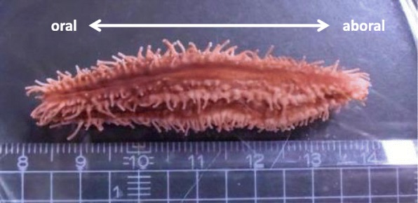
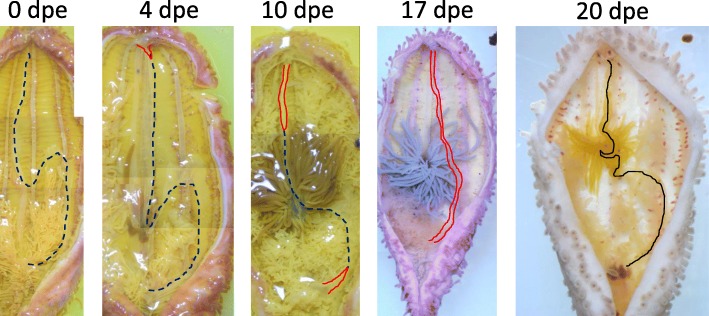
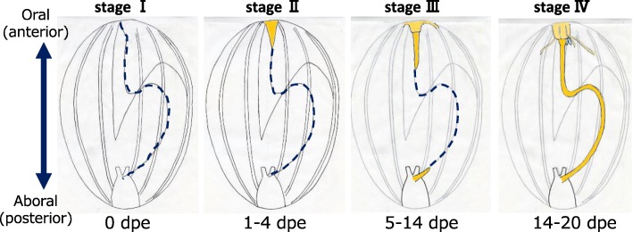
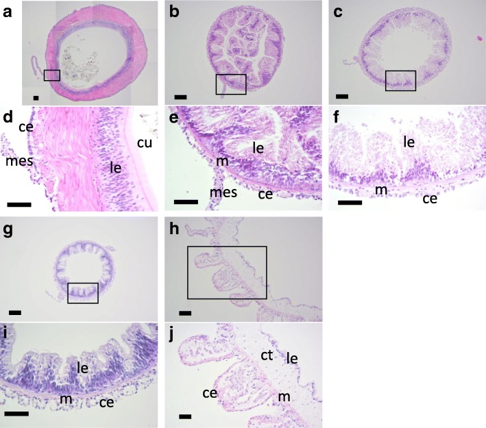
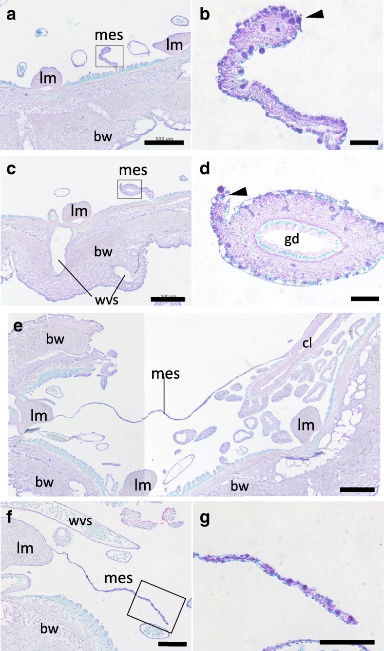
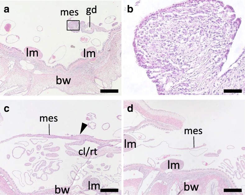
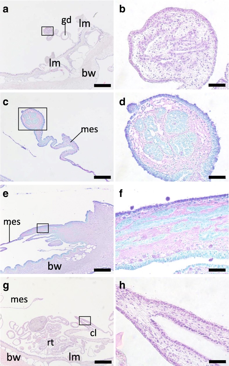
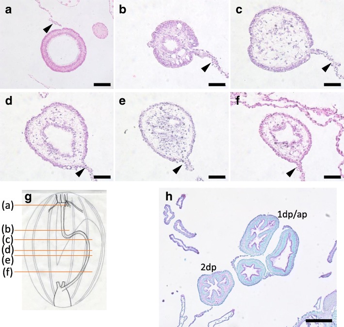
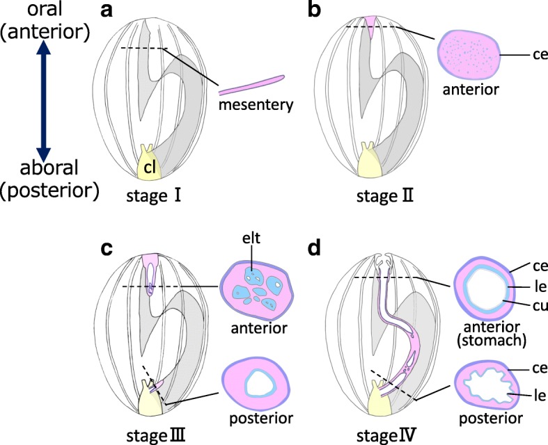
Sea cucumbers (a class of echinoderms) exhibit a high capacity for regeneration, such that, following ejection of inner organs in a process called evisceration, the lost organs regenerate. There are two ways by which evisceration occurs in sea cucmber species: from the mouth (anterior) or the anus (posterior). Intriguingly, regenerating tissues are formed at both the anterior and posterior regions and extend toward the opposite ends, and merge to form a complete digestive tract. From the posterior side, the digestive tube regenerates extending a continuous tube from the cloaca, which remains at evisceration. In posteriorly-eviscerating species, the esophagus remains in the body, and a new tube regenerates continuously from it. However, in anterior-eviscerating species, no tubular tissue remains in the anterior region, raising the question of how the new digestive tube forms in the anterior regenerate. We addressed this question by detailed histological observations of the regenerating anterior digestive tract in a small sea cucumber, Eupentacta quinquesemita ("ishiko" in Japanese) after induced-evisceration. We found that an initial rudiment consisting of mesenchymal cells is formed along the edge of the anterior mesentery from the anterior end, and then, among the mesenchymal cells, multiple clusters of epithelial-like cells appears simultaneously and repeatedly in the extending region by mesenchymal-epithelial transition (MET) as visulalized using toluidine blue staining. Subsequently, multiple cavities were formed surrounded with these epithelial cells, and appeared to coalesce with each other to form into multiple lumens, and to eventually become a single tube. This anterior tube then fused to the tube regenerated from the posterior rudiment. Thus, we elucidated the process of regeneration of the anterior portion of the gut in an anteriorly eviscerating species, and suggest the involvement of MET and fusion of cavities/lumens in regeneration of the digestive tube.

Keywords: Digestive tract; Echinoderm; Evisceration; Mesenchymal–epithelial transition; Mesentery; Regeneration; Sea cucumber.


Conflict of interest statement

Competing interestsThe authors declare that they have no competing interests.

Figures

Fig. 1
Fig. 1
The animal used in the study. A small sea cucumber, Eupentacta quinquesemita, is about 5 cm in size. The left side in this figure is oral (anterior), and the right side is aboral (posterior)
Fig. 2
Fig. 2
Internal morphology of regenerating animals. Animals just after evisceration (0 dpe) to 20 dpe were dissected along the body length, at the right side of the mid-ventral ambulacrum and flattened. There are slight color variations of the animals, but specimens at 0, 4, and 10 dpe were fixed with Bouin’s fixative and thus appear yellowish. The animals were about 4–5 cm in length. Black broken line: the edge of mesentery; red solid line: area along the edge of the mesentery, with thickened gut rudiments; black line: grown gut rudiment observable without the microscope
Fig. 3
Fig. 3
Stages of regeneration. Based on the internal morphology of regenerating animals, we defined four stages of regeneration, stages I to IV, and schematic diagrams of each are shown. Blue broken line: the edge of mesentery without regenerating tissues; yellow colored area: regenerating tissues (gut rudiment)
Fig. 4
Fig. 4
Histology of the intact digestive tract. Cross sections of the digestive tract of E. quinquesemita were stained with HE. d, e, f, i, j are high-magnification views of the boxed areas in a, b, c, g, h, respectively. a, d Stomach. Some contents of the stomach are also observed in a. b, e 1st descending intestine. c, f Ascending intestine. g, i 2nd descending intestine. h, j Cloaca. ce, celomic epithelium; ct, connective tissue; cu, cuticle; gd, gonadal duct; le, luminal epithelium; m, muscle; mes, mesentery. Scale bars100 μm in a-c, g, h; 50 μm in d-f, i, j
Fig. 5
Fig. 5
Histology of the tissues at stage I. Cross sections of the body at stage I (0 dpe) were stained with TB, mainly focusing on the anterior (a-d) and posterior (e, f) mesenteries. b, d and g are high-magnification views of the boxed areas in a, c and f, respectively. a, b Anterior tissue, posterior to the anterior rudiment. c, d Anterior tissue posterior than a and b. The gonadal duct remains in the mesentery, but no structures are present at the free end. e Posterior tissue. The cloaca and the body wall are connected by the mesentery. f, g Posterior tissue, anterior than e. In b, d and g, no tubular structures nor developed cell mass are observed at the free edge of the mesentery (arrowhead). bw, body wall; cl, cloaca; gd, gonadal duct; lm, longitudinal muscle; mes, mesentery; wvs: a part of the water vascular system (radial water canals, etc.). Arrowheads indicate the free edges of the mesentery. Scale bars 500 μm in a, c, e; 200 μm in f; 50 μm in b, d, g
Fig. 6
Fig. 6
Histology of the tissues at stage II. Cross sections of the body at stage II (4 dpe) were stained with HE, focusing on the mesentery at the anterior (a, b) and posterior (c, d) regions of the body. a, b Anterior mesentery. A mass of cells is present on the free edge of the mesentery. b is a high-magnification view of the boxed area in a. c Posterior tissue. A thickening of tissue (arrowhead) is observed at the connection of the mesentery and the cloaca. Respiratory trees are an organ continuous from the cloaca, and thus these two tissues could not be strictly distinguished. d Posterior tissue, anterior than c. A part of the mesentery with a free edge is shown. bw, body wall; cl/rt., cloaca or respiratory trees; gd, gonadal duct; lm, longitudinal muscle; mes, mesentery. Scale bars 500 μm in a, c, d; 50 μm in b
Fig. 7
Fig. 7
Histology of the tissues at stage III. Histological sections of the anterior (a-f, i) and posterior (g, h) regions of the body at stage III. b, d, f, h are high-magnification views of the boxed areas in a, c, e, g, respectively. a, b Cross section, 6 dpe, stained with HE. c, d Cross section, 7 dpe, stained with TB. e, f Longitudinal section of the oral region, 7 dpe, stained with TB. The right side of this figure is anterior. g, h Posterior region, 6 dpe, stained with HE. The animal was sliced perpendicular to the oral-aboral axis but a longitudinal section of the regenerating tube was made. In the lumen, a mass of obscure material is observed (arrowhead). bw, body wall; cl, cloaca; gd, gonadal duct; mes, mesentery; rt., respiratory trees. Scale bar 500 μm in a, e, g; 200 μm in c; 50 μm in b, d, h, f
Fig. 8
Fig. 8
Histology of the tissues at stage IV. Cross sections of the body at stage IV focusing on the regenerated digestive tract. Sections a-f were made at positions indicated in g, of a specimen at 17 dpe. h The middle part of the animal with 1st descending, ascending and 2nd descending intestines, at 20 dpe, stained with TB. The slice was made roughly between b and c in a further regenerated animal with a single tube throughout the digestive tract. The 1st descending and ascending intestines could not be distinguished, since the intestines meandered. 1st descending or ascending intestine; 2dp, 2nd descending intestine. Arrowheads indicate mesenteries. Scale bar 50 μm in b-f; 100 μm in a; 200 μm in h
Fig. 9
Fig. 9
Schematic diagram of regeneration of the digestive tube. The internal structure of dissected animals and cross sections at the positions of the dotted lines are illustrated. Organs other than the mesentery and the digestive tract are ommited. a Stage I, just after evisceration. Only the mesentery (gray shaded area) and the cloaca (cl; yellow) remain in the body cavity. b Stage II. A regenerating tissue (gut rudiment) appears at the anterior side as a mass of mainly mesenchymal cells surrounded by coelomic epithelium. c Stage III. Multiple cavities are formed in the anterior regenerating tissue and these coalesce with each other to form lumens. d At stage IV when regeneration is more progressed, the gut rudiment (thickened tissue on the mesentery) becomes continuous between the anterior and posterior sides. The figure shows that the anterior and posterior lumens are not yet connected, but later a single continuous tube is completed. Intestines differentiate according to their position in the digestive tract. In the stomach, a muscle layer (not shown) develops and luminal epithelium (le) is covered with cuticles (cu), as in intact tissues (see Fig. 4a, d). ce: coelomic epithelium; cl: cloaca; cu: cuticles; elt: epithelium-like tissue; le: luminal epithelum

Similar articles

Cited by

References

    1. Tanaka EM, Reddien PW. The cellular basis for animal regeneration. Dev Cell. 2011;21:172–185. doi: 10.1016/j.devcel.2011.06.016. - DOI - PMC - PubMed
    1. Grillo M, Konstantinides N, Averof M. Old questions, new models: unraveling complex organ regeneration with new experimental approaches. Curr Opin Genet Dev. 2016;40:23–31. doi: 10.1016/j.gde.2016.05.006. - DOI - PubMed
    1. Arnone MI, Byrne M, Martinez P. Echinodermata. In: Wanninger A, editor. Evolutionnary developmental biology of invertebrates: 6 Deuterostomia. 2015. pp. 1–58.
    1. Ferrario C, Ben Khadra Y, Czarkwiani A, Zakrzewski A, Martinez P, Colombo G, et al. Fundamental aspects of arm repair phase in two echinoderm models. Dev Biol. 2018;433:297–309. doi: 10.1016/j.ydbio.2017.09.035. - DOI - PMC - PubMed
    1. Candia Carnevali MD, Bonasoro F. Microscopic overview of crinoid regeneration. Microsc Res Tech. 2001;55:403–426. doi: 10.1002/jemt.1187. - DOI - PubMed