. 2019 Jul 5:5:113.
doi: 10.1038/s41420-019-0192-4. eCollection 2019.

24(S)-Hydroxycholesterol induces ER dysfunction-mediated unconventional cell death

Affiliations

24(S)-Hydroxycholesterol induces ER dysfunction-mediated unconventional cell death

Yasuomi Urano et al. Cell Death Discov. .

Abstract

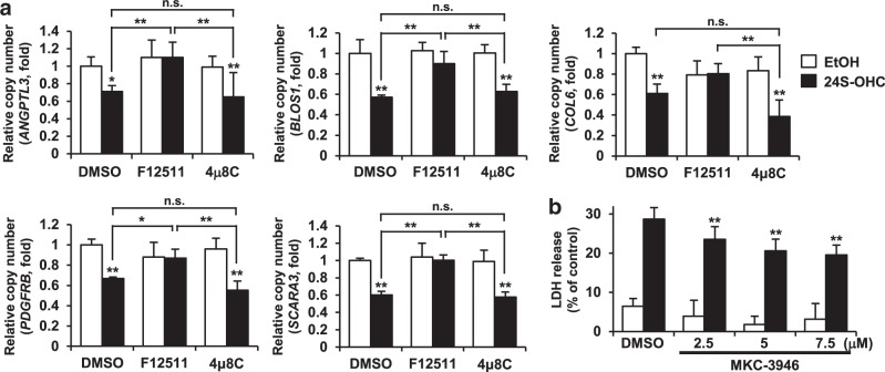
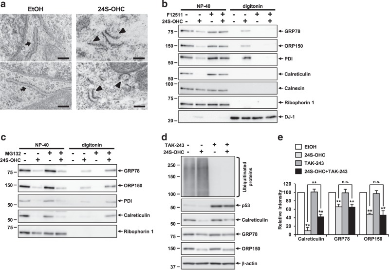
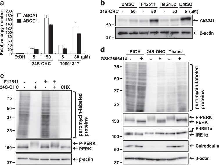
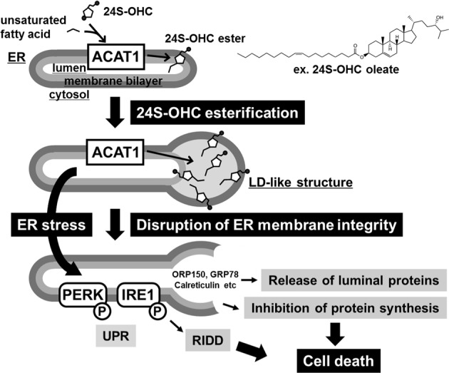
Endoplasmic reticulum (ER) stress induced by disruption of protein folding activates the unfolded protein response (UPR), which while generally pro-survival in effect can also induce cell death under severe ER stress. 24(S)-hydroxycholesterol (24S-OHC), which is enzymatically produced in the ER of neurons, plays an important role in maintaining brain cholesterol homeostasis but also shows neurotoxicity when subjected to esterification by acyl-CoA:cholesterol acyltransferase 1 (ACAT1) in the ER. In this study, we demonstrated that the accumulation of 24S-OHC esters in human neuroblastoma SH-SY5Y cells evoked the UPR with substantially no pro-survival adaptive response but with significant activation of pro-death UPR signaling via regulated IRE1-dependent decay (RIDD). We further found that accumulation of 24S-OHC esters caused disruption of ER membrane integrity and release of ER luminal proteins into cytosol. We also found that de novo synthesis of global proteins was robustly suppressed in 24S-OHC-treated cells. Collectively, these results show that ER dysfunction and the accompanying RIDD-mediated pro-death UPR signaling and global protein synthesis inhibition are responsible for 24S-OHC ester-induced unconventional cell death.

Keywords: Apoptosis; Sterols.


Conflict of interest statement

Conflict of interestThe authors declare that they have no conflict of interest.

Figures

Fig. 1
Fig. 1. Accumulation of 24S-OHC esters activated the UPR signaling pathway and downregulated expression of ER chaperone proteins in SH-SY5Y cells.
ac SH-SY5Y cells were pretreated with 5 μM F12511 for 15 min or with 100 μM Nec-1 (a) for 1 h and then exposed to 50 μM 24S-OHC for 6 h. Cells were also treated with 3 μM thapsigargin (Thapsi) for 3 h. a Whole-cell lysates were subjected to immunoblotting with appropriate antibodies as indicated. b The unspliced (XBP1u) and spliced (XBP1s) XBP1 mRNAs were analyzed by RT-PCR. c Whole-cell lysates were immunoblotted with antibodies specific for XBP1s or β-actin. d Cells were pretreated with 20 μM MG132 for 30 min and then exposed to 50 μM 24S-OHC or 3 μM thapsigargin for 6 h. Whole-cell lysates were immunoblotted with antibodies specific for ATF6 or β-actin. Asterisks denote nonspecific bands. e, f Cells were treated as in panel a. g, h Cells were treated as in panel d. e, g Whole-cell lysates were subjected to immunoblotting with appropriate antibodies as indicated. f, h Band intensities were quantified by densitometric scanning, relative intensity is shown. Mean ± SD n = 3, **P < 0.01, n.s. not significant
Fig. 2
Fig. 2. PERK and IRE1-XBP1 axes were not involved in 24S-OHC-induced cell death in SH-SY5Y cells.
a SH-SY5Y cells were treated as in Fig. 1a. Whole-cell lysates were immunoblotted with antibodies specific for ATF4, CHOP, or β-actin. b, c Cells were transfected with CHOP (siCHOP #1-#3) or negative control (NC) siRNA oligo for 24 h. b Whole-cell lysates were immunoblotted with antibodies specific for CHOP or β-actin. Relative expression levels of CHOP are shown. c The cells were exposed to 50 μM 24S-OHC for 24 h. Cell viability was measured by LDH assay. **P < 0.01, when compared with cells treated with vehicle or among 24S-OHC-treated groups. d Cells were pretreated with 5 μM GSK2606414 for 30 min then exposed to 50 μM 24S-OHC or 3 μM thapsigargin for 6 h. Whole-cell lysates were immunoblotted with antibodies specific for PERK, IRE1α, or β-actin. e Cells were pretreated with 5 or 10 μM GSK2606414 for 30 min then exposed to 50 μM 24S-OHC for 24 h. Cell viability was measured by WST-8 assay. **P < 0.01. f Cells were pretreated with 5 μM F12511 for 15 min or with 1–10 μM 4μ8 C for 1 h then exposed to 50 μM 24S-OHC for 6 h or 1 μM thapsigargin for 3 h, respectively. XBP1 mRNAs were analyzed by RT-PCR. g Cells were pretreated with 3 or 10 μM 4μ8 C for 1 h and then exposed to 50 μM 24S-OHC for 24 h. Cell viability was measured by WST-8 assay. **P < 0.01
Fig. 3
Fig. 3. Inhibition of RIDD mitigated 24S-OHC-induced cell death in SH-SY5Y cells.
a SH-SY5Y cells were pretreated with 5 μM F12511 for 15 min or with 10 μM 4μ8 C for 1 h and then exposed to 50 μM 24S-OHC for 6 h. The mRNA levels of RIDD target genes (ANGPTL3, BLOS1, COL6, PDGFRB, and SCARA3) were quantified by real-time PCR. Results are shown as normalized to levels measured for RPL32. *P < 0.05, **P < 0.01, when compared with cells treated with vehicle or among 24S-OHC-treated groups. b Cells were pretreated with 2.5–7.5 μM MKC-3946 for 1 h then exposed to 50 μM 24S-OHC for 24 h. Cell viability was measured by LDH assay. **P < 0.01, when compared with cells treated with 24S-OHC alone
Fig. 4
Fig. 4. Accumulation of 24S-OHC esters induced disruption of ER membrane integrity, resulting in release of ER luminal proteins into cytosol, in SH-SY5Y cells.
a SH-SY5Y cells were treated with 50 μM 24S-OHC for 3 h. Cells were subjected to electron microscopy. Representative images are shown. Arrows indicate intact rough ER. Arrowheads indicate broken-membrane ER structures. Scale bar, 0.2 μm. b, c Cells were pretreated with 5 μM F12511 for 15 min (b) or 20 μM MG132 for 30 min (c) and then exposed to 50 μM 24S-OHC for 3 h (b) or 6 h (c). Cells were then subjected to sequential detergent extraction using digitonin and NP-40 as indicated. Equal aliquots from each fraction were immunoblotted using antibodies as indicated. d, e Cells were pretreated with 5 μM TAK-243 for 30 min and then exposed to 50 μM 24S-OHC for 6 h. d Whole-cell lysates were subjected to immunoblotting with appropriate antibodies as indicated. e Band intensities were quantified by densitometric scanning, relative intensity being shown. n = 3, **P < 0.01, when compared with cells treated with vehicle or among 24S-OHC-treated groups
Fig. 5
Fig. 5. Global protein synthesis was inhibited by 24S-OHC in SH-SY5Y cells.
a SH-SY5Y cells were treated with 24S-OHC (5 or 50 μM) or T0901317 (5 or 80 μM) for 6 h. The mRNA levels of ABCA1 and ABCG1 were quantified by real-time PCR. Results are shown as normalized to levels measured for RPL32. b Cells were pretreated with 5 μM F12511 or 5 μM MG132 for 30 min and then exposed to 5 or 50 μM 24S-OHC for 6 h. Whole-cell lysates were immunoblotted with antibodies specific for ABCG1 or β-actin. c Cells were pretreated with or without 5 μM F12511 for 15 min and then exposed to 50 μM 24S-OHC or 10 μg/ml CHX for 6 h. d Cells were pretreated with 5 μM GSK2606414 for 30 min and then exposed to 50 μM 24S-OHC or 3 μM thapsigargin for 6 h. c, d Cells were then incubated with 10 μg/ml puromycin for last 15 min. Whole-cell lysates were immunoblotted with antibodies specific for puromycin, PERK, IRE1α, calreticulin, or β-actin
Fig. 6
Fig. 6. Schematic of proposed mechanism for 24S-OHC-induced cell death in SH-SY5Y cells.
Under conditions of excess 24S-OHC in SH-SY5Y cells, ACAT1 catalyzes 24S-OHC ester formation from 24S-OHC and long-chain unsaturated fatty acid in the ER. Accumulation of 24S-OHC esters between the two leaflets of the ER membrane bilayer leads to formation of an LD-like structure coupled with a swollen ER structure, resulting in disruption of ER membrane integrity, which in turn induces release of ER luminal proteins. ER stress and/or disruption of ER membrane integrity activates pro-death UPR signaling including RIDD and causes inhibition of protein synthesis, triggering one unconventional type of cell death

Similar articles

Cited by

References

    1. Hetz C. The unfolded protein response: controlling cell fate decisions under ER stress and beyond. Nat. Rev. Mol. Cell. Biol. 2012;13:89–102. doi: 10.1038/nrm3270. - DOI - PubMed
    1. Moore KA, Hollien J. The unfolded protein response in secretory cell function. Annu. Rev. Genet. 2012;46:165–183. doi: 10.1146/annurev-genet-110711-155644. - DOI - PubMed
    1. Kim I, Xu W, Reed JC. Cell death and endoplasmic reticulum stress: disease relevance and therapeutic opportunities. Nat. Rev. Drug Discov. 2008;7:1013–1030. doi: 10.1038/nrd2755. - DOI - PubMed
    1. Tabas I, Ron D. Integrating the mechanisms of apoptosis induced by endoplasmic reticulum stress. Nat. Cell Biol. 2011;13:184–190. doi: 10.1038/ncb0311-184. - DOI - PMC - PubMed
    1. Sano R, Reed JC. ER stress-induced cell death mechanisms. Biochim. Biophys. Acta. 2013;1833:3460–3470. doi: 10.1016/j.bbamcr.2013.06.028. - DOI - PMC - PubMed