

Wide phenotypic variability in RSPH9-associated primary ciliary dyskinesia: review of a case-series from Cyprus
- PMID: 31285900
- PMCID: PMC6588774
- DOI: 10.21037/jtd.2019.04.71
Wide phenotypic variability in RSPH9-associated primary ciliary dyskinesia: review of a case-series from Cyprus
Abstract
Background: Primary ciliary dyskinesia (PCD) is an inherited ciliary motility disorder caused by mutations in at least 40 genes. RSPH9 gene mutations encoding aberrant radial spoke head proteins have been linked with PCD. The clinical spectrum extent of RSPH9 gene mutations remains to date largely unknown. We aimed to describe the diagnostic and clinical phenotype in a case-series of RSPH9-associated PCD.
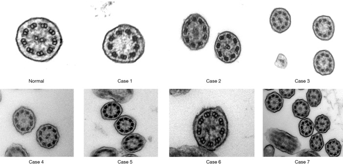
Methods: We performed whole exome sequencing in suspect patients from Cyprus who on repeated cilia biopsies demonstrated loss of the central pair apparatus on Transmission Electron Microscopy (TEM) and rotary beating patterns on High Speed Video Microscopy (HSVM), compatible to findings described previously in PCD patients bearing pathogenic RSPH9 mutations. In cases confirmed by genetic testing, we reviewed diagnostic, demographic and clinical data, as well as anthropometric and spirometric measurements.
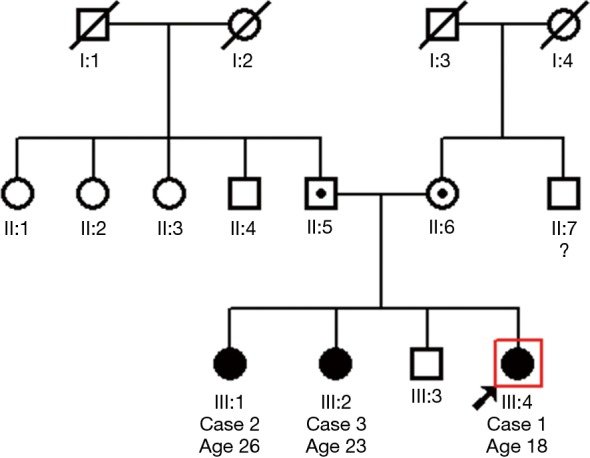
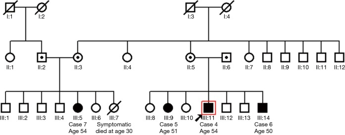
Results: We diagnosed 7 individuals (5 females) homozygous for the novel RSPH9 splice site mutation c.670+2T>C in intron 4, who originated from two families. Despite bearing the same genetic variant, patients presented a highly variable age (median 47.9 years; range, 6.6 to 51.4 years) and with a diverse clinical picture, all reporting a history of chronic or recurrent wet cough (100%), and at varying frequencies neonatal respiratory distress (43%), chronic rhinosinusitis (71%), and wheezing (43%). Complications such as bronchiectasis (71%), history of pneumonia(s) (57%) and surgical interventions (43%) clustered in some patients displaying typical PCD, but not in others with milder phenotypes. BMI-z scores (median: 0.53; range, -0.69 to 1.52), FEV1-z scores (median: -0.37; range: -1.79 to 0.22) and FVC z-scores (median: -0.80; range: -2.01 to 0.36) were on average within the normal range, although slightly reduced.
Conclusions: In conclusion, RSPH9-associated PCD disease demonstrates wide phenotypic variability. In some cases, mild clinical presentation is difficult to justify diagnostic work-up, highlighting the importance of wider adoption of genetic diagnostics. Larger studies are needed to assess variability of clinical spectrum associated to alterations of PCD genes.
Keywords: Ciliary motility disorders; Kartagener syndrome; bronchiectasis.
Conflict of interest statement
Conflicts of Interest: The authors have no conflicts of interest to declare.
Figures


Similar articles
-
Mutations in radial spoke head genes and ultrastructural cilia defects in East-European cohort of primary ciliary dyskinesia patients.PLoS One. 2012;7(3):e33667. doi: 10.1371/journal.pone.0033667. Epub 2012 Mar 20. PLoS One. 2012. PMID: 22448264 Free PMC article.
-
Mapping the Most Common Founder Variant in RSPH9 That Causes Primary Ciliary Dyskinesia in Multiple Consanguineous Families of Bedouin Arabs.J Clin Med. 2023 Oct 13;12(20):6505. doi: 10.3390/jcm12206505. J Clin Med. 2023. PMID: 37892643 Free PMC article.
-
Mutations in RSPH1 cause primary ciliary dyskinesia with a unique clinical and ciliary phenotype.Am J Respir Crit Care Med. 2014 Mar 15;189(6):707-17. doi: 10.1164/rccm.201311-2047OC. Am J Respir Crit Care Med. 2014. PMID: 24568568 Free PMC article.
-
Value of transmission electron microscopy for primary ciliary dyskinesia diagnosis in the era of molecular medicine: Genetic defects with normal and non-diagnostic ciliary ultrastructure.Ultrastruct Pathol. 2017 Nov-Dec;41(6):373-385. doi: 10.1080/01913123.2017.1362088. Epub 2017 Sep 15. Ultrastruct Pathol. 2017. PMID: 28915070 Free PMC article. Review.
-
[Cilia ultrastructural and gene variation of primary ciliary dyskinesia: report of three cases and literatures review].Zhonghua Er Ke Za Zhi. 2018 Feb 2;56(2):134-137. doi: 10.3760/cma.j.issn.0578-1310.2018.02.012. Zhonghua Er Ke Za Zhi. 2018. PMID: 29429202 Review. Chinese.
Cited by
-
Nasal nitric oxide measurement for primary ciliary dyskinesia diagnosis: The impact of underlying genetic defects on diagnostic accuracy.Pediatr Investig. 2019 Dec 21;3(4):214-216. doi: 10.1002/ped4.12171. eCollection 2019 Dec. Pediatr Investig. 2019. PMID: 32851325 Free PMC article. No abstract available.
-
First reports of primary ciliary dyskinesia caused by a shared DNAH11 allele in Canadian Inuit.Pediatr Pulmonol. 2023 Jul;58(7):1942-1949. doi: 10.1002/ppul.26414. Epub 2023 Apr 23. Pediatr Pulmonol. 2023. PMID: 37088965 Free PMC article.
-
Observational study of health utilities in adult primary ciliary dyskinesia patients: preliminary data on associations with molecular diagnosis, clinical phenotype and HRQOL measures.Multidiscip Respir Med. 2022 Dec 20;17:881. doi: 10.4081/mrm.2022.881. eCollection 2022 Jan 12. Multidiscip Respir Med. 2022. PMID: 36636646 Free PMC article.
-
The impact of primary ciliary dyskinesia on female and male fertility: a narrative review.Hum Reprod Update. 2023 May 2;29(3):347-367. doi: 10.1093/humupd/dmad003. Hum Reprod Update. 2023. PMID: 36721921 Free PMC article. Review.
-
Emerging Genotype-Phenotype Relationships in Primary Ciliary Dyskinesia.Int J Mol Sci. 2021 Jul 31;22(15):8272. doi: 10.3390/ijms22158272. Int J Mol Sci. 2021. PMID: 34361034 Free PMC article. Review.
References
LinkOut - more resources
Full Text Sources