

Comparing the diagnostic value of 18F-FDG-PET/CT versus CT for differentiating benign and malignant solitary pulmonary nodules: a meta-analysis
- PMID: 31285902
- PMCID: PMC6588752
- DOI: 10.21037/jtd.2019.05.21
Comparing the diagnostic value of 18F-FDG-PET/CT versus CT for differentiating benign and malignant solitary pulmonary nodules: a meta-analysis
Abstract
Background: This quantitative meta-analysis was conducted to provide an indirect comparison of the diagnostic value of computed tomography (CT) with positron emission tomography (PET)/CT for differentiating benign and malignant solitary pulmonary nodules (SPNs).
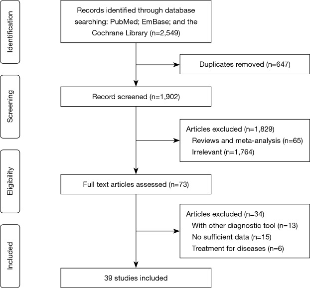
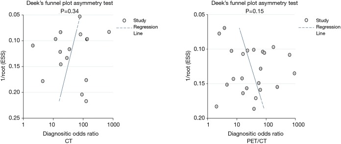
Methods: PubMed, Embase, and the Cochrane Library were searched to identify eligible studies throughout November 2018, which differentiated benign and malignant SPNs using CT or PET/CT. The summary sensitivity, specificity, positive and negative likelihood ratio (PLR and NLR), diagnostic odds ratio (DOR), and area under the receiver operating characteristic curve (AUC) were calculated using bivariate generalized linear mixed model and random-effects model. The diagnostic value of CT with PET/CT was indirectly evaluated using the ratio for diagnostic parameters.
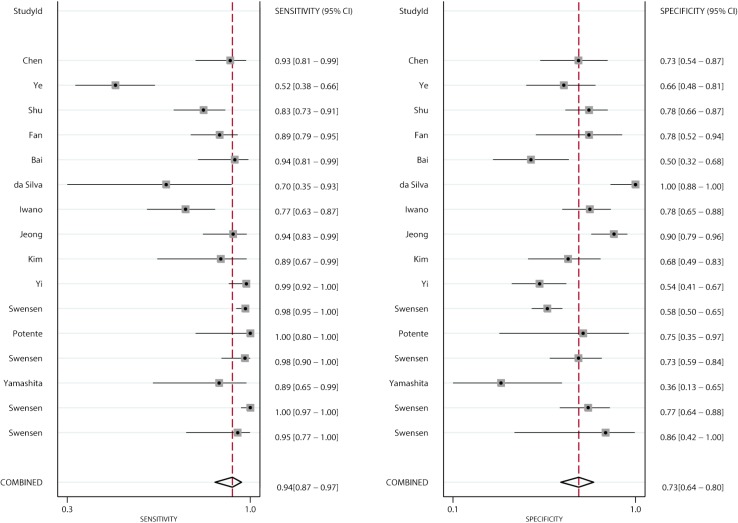
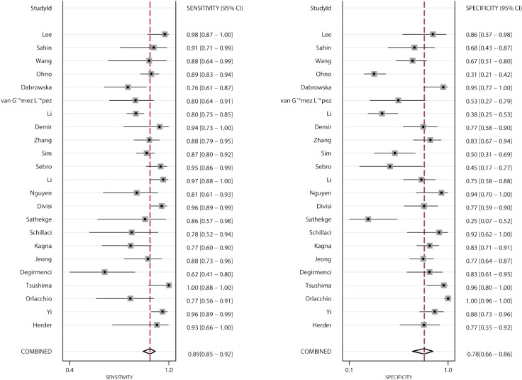
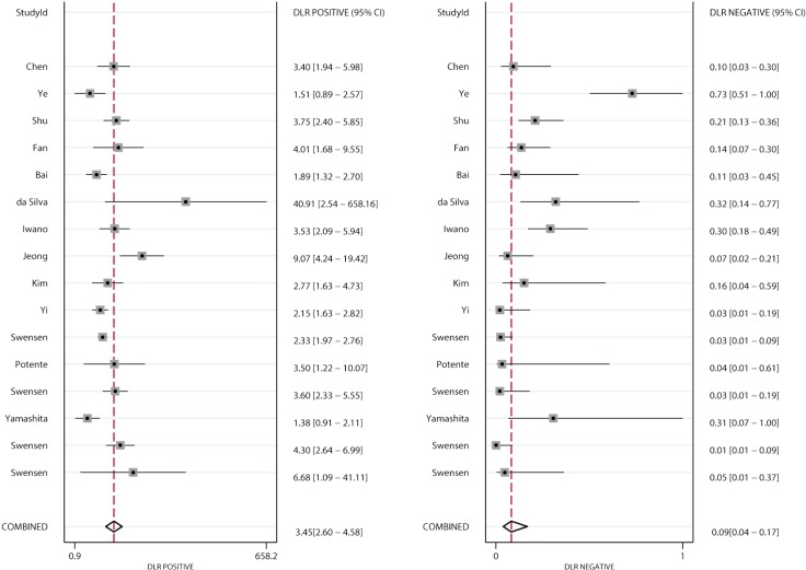
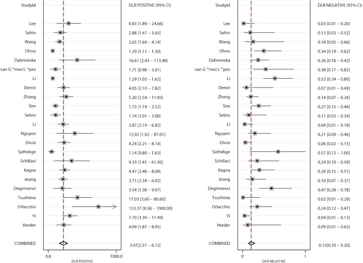
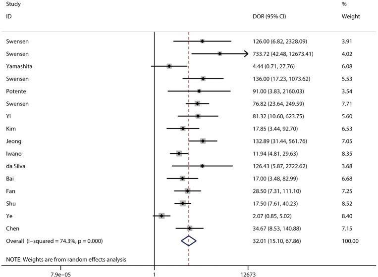
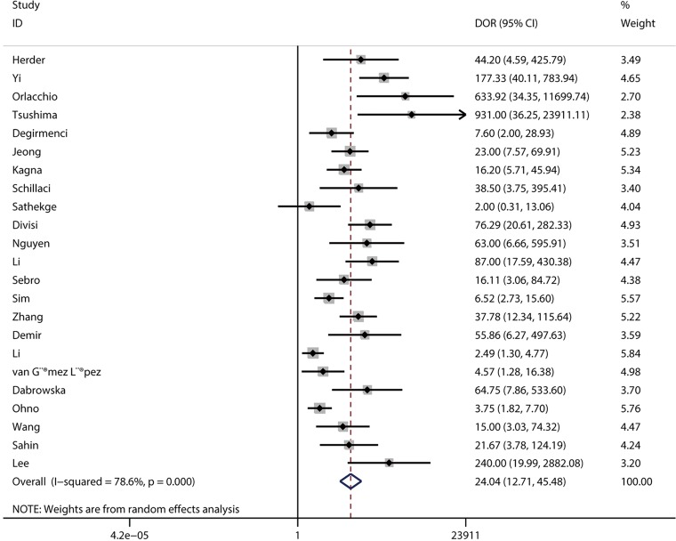
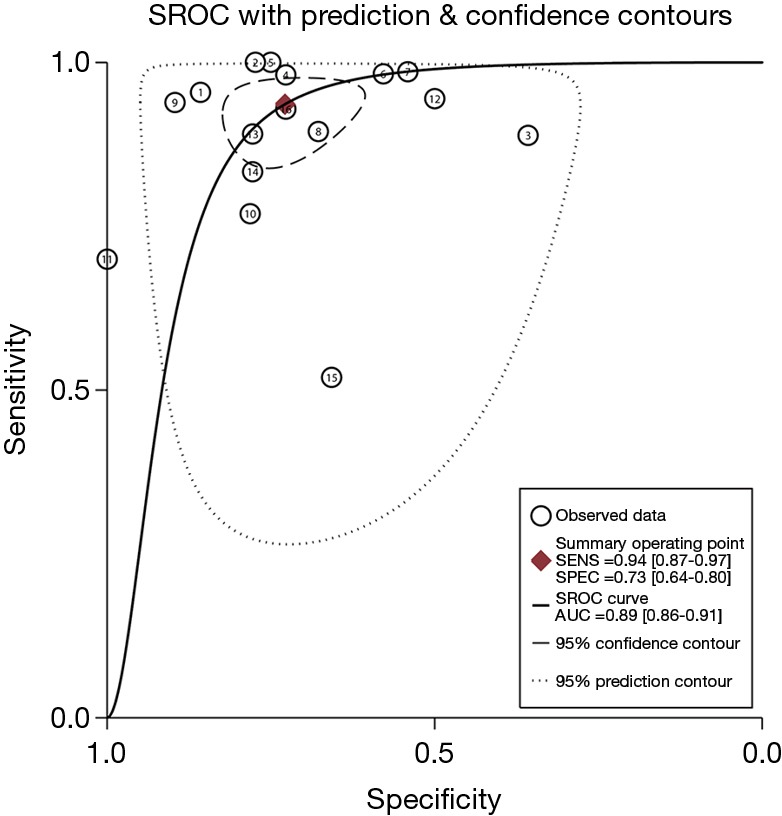
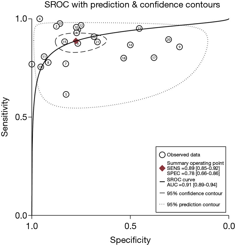
Results: The sensitivity, specificity, PLR, NLR, DOR, and AUC for CT were 0.94 [95% confidence interval (CI): 0.87-0.97], 0.73 (95% CI: 0.64-0.80), 3.45 (95% CI: 2.60-4.58), 0.09 (95% CI: 0.04-0.17), 32.01 (95% CI: 15.10-67.86), and 0.89 (95% CI: 0.86-0.91), respectively. The pooled sensitivity, specificity, PLR, NLR, DOR, and AUC for PET/CT were 0.89 (95% CI: 0.85-0.92), 0.78 (95% CI: 0.66-0.86), 3.97 (95% CI: 2.57-6.13), 0.15 (95% CI: 0.10-0.20), 24.04 (95% CI: 12.71-45.48), and 0.91 (95% CI: 0.89-0.94), respectively. No significant differences were observed between CT and PET/CT for sensitivity, specificity, PLR, NLR, DOR, and AUC.
Conclusions: This study used both CT and PET/CT with a moderate-to-high diagnostic value for differentiating benign and malignant SPNs and showed no significant differences in diagnostic parameters between CT and PET/CT.
Keywords: 18F-FDG-PET/CT; benign solitary pulmonary nodules (benign SPNs); diagnosis; malignant solitary pulmonary nodules (malignant SPNs).
Conflict of interest statement
Conflicts of Interest: The authors have no conflicts of interest to declare.
Figures
References
LinkOut - more resources
Full Text Sources