

Decontamination methods to restore the biocompatibility of contaminated titanium surfaces
- PMID: 31285943
- PMCID: PMC6599751
- DOI: 10.5051/jpis.2019.49.3.193
Decontamination methods to restore the biocompatibility of contaminated titanium surfaces
Abstract
Purpose: The reaction of cells to a titanium implant depends on the surface characteristics of the implant which are affected by decontamination. The aim of this study was to evaluate the cytocompatibility of titanium disks treated with various decontamination methods, using salivary bacterial contamination with dental pellicle formation as an in vitro model.
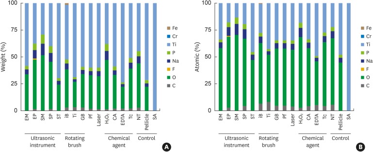
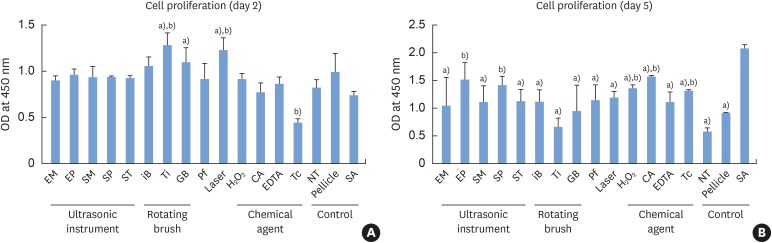
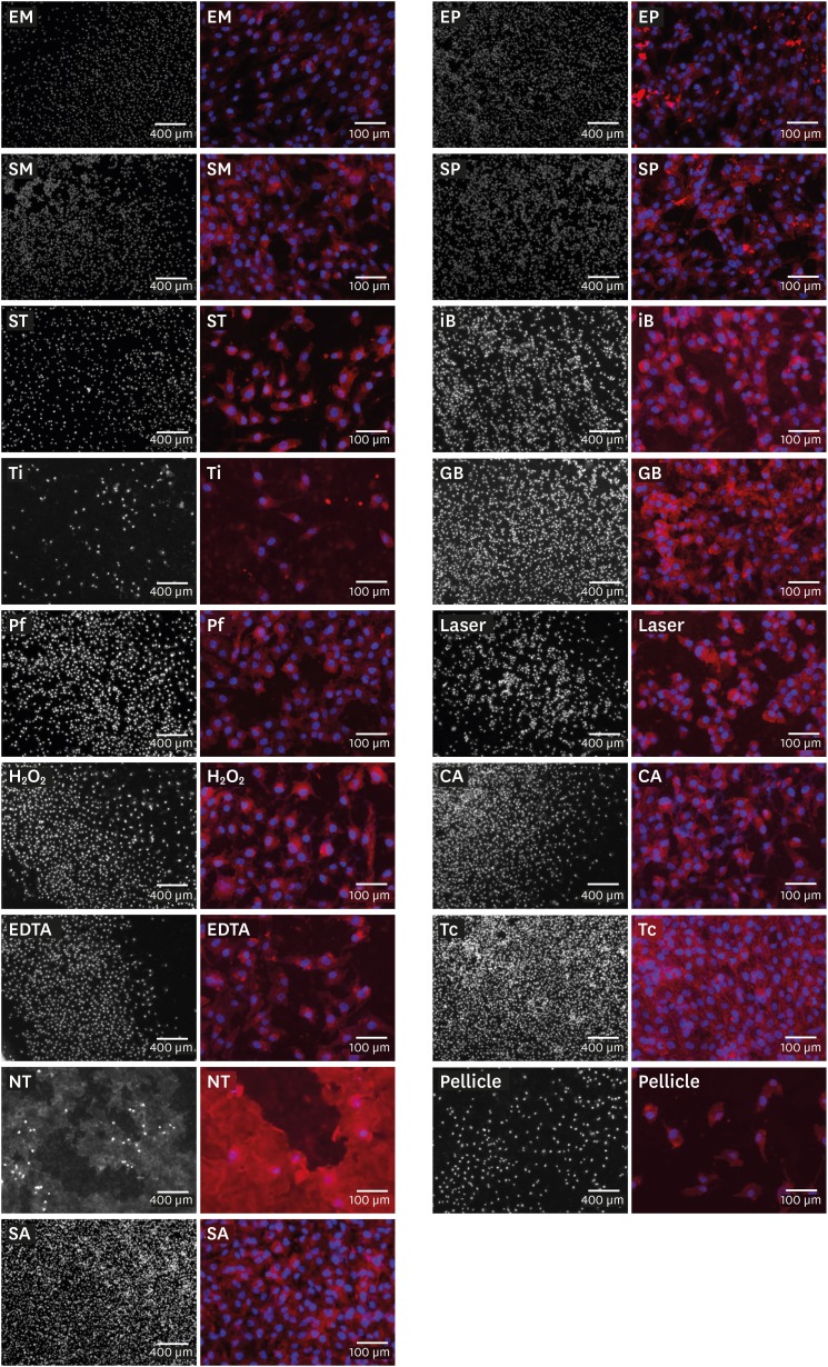
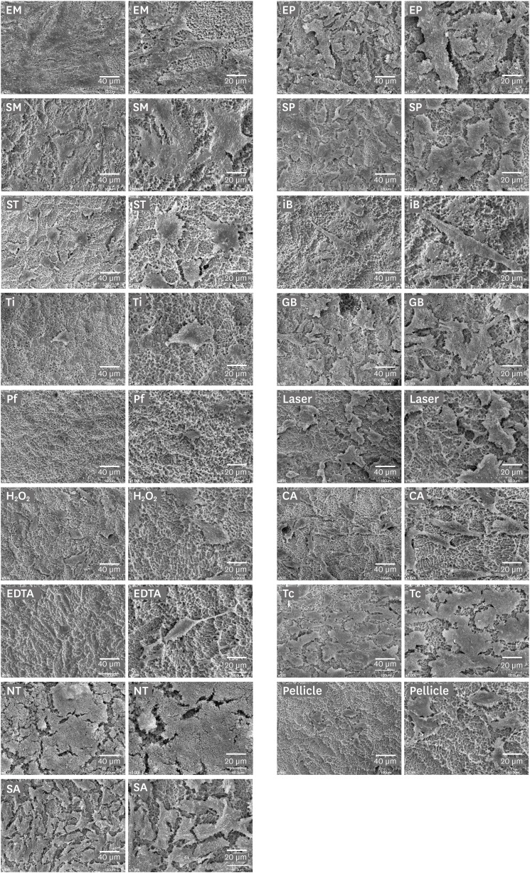
Methods: Sand-blasted and acid-etched (SA) titanium disks were used. Three control groups (pristine SA disks [SA group]; salivary pellicle-coated SA disks [pellicle group]; and biofilm-coated, untreated SA disks [NT group]) were not subjected to any decontamination treatments. Decontamination of the biofilm-coated disks was performed by 14 methods, including ultrasonic instruments, rotating instruments, an air-powder abrasive system, a laser, and chemical agents. MG63 cells were cultured in the presence of the treated disks. Cell proliferation assays were performed on days 2 and 5 of cell culture, and cell morphology was analyzed by immunofluorescence and scanning electron microscopy (SEM). A vascular endothelial growth factor (VEGF) assay was performed on day 5 of culture.
Results: The cell proliferation assay revealed that all decontaminated disks, except for the 2 groups treated using a plastic tip, showed significantly less cell proliferation than the SA group. The immunofluorescence and SEM analyses revealed that most groups showed comparable cell density, with the exception of the NT group, in which the cell density was lower and bacterial residue was observed. Furthermore, the cells grown with tetracycline-treated titanium disks showed significantly lower VEGF production than those in the SA group.
Conclusions: None of the decontamination methods resulted in cytocompatibility similar to that of pristine SA titanium. However, many methods caused improvement in the biocompatibility of the titanium disks in comparison with the biofilm-coated, untreated titanium disks. This suggests that decontamination is indispensable for the treatment of peri-implantitis, even if the original biocompatibility cannot be restored.
Keywords: Biocompatible materials; Decontamination; Dental implants; Peri-implantitis.
Conflict of interest statement
Conflict of interest: No potential conflict of interest relevant to this article was reported.
Figures

Similar articles
-
The Effect of NiTi Brush, Polishing Brush, and Chemical Agent on the Dental Implant Surface Morphology and Cytocompatibility.Clin Implant Dent Relat Res. 2025 Feb;27(1):e13417. doi: 10.1111/cid.13417. Epub 2024 Nov 21. Clin Implant Dent Relat Res. 2025. PMID: 39569703 Free PMC article.
-
Antimicrobial Agents Used in the Treatment of Peri-Implantitis Alter the Physicochemistry and Cytocompatibility of Titanium Surfaces.J Periodontol. 2016 Jul;87(7):809-19. doi: 10.1902/jop.2016.150684. Epub 2016 Feb 28. J Periodontol. 2016. PMID: 26923474
-
The Effect of Microcosm Biofilm Decontamination on Surface Topography, Chemistry, and Biocompatibility Dynamics of Implant Titanium Surfaces.Int J Mol Sci. 2022 Sep 2;23(17):10033. doi: 10.3390/ijms231710033. Int J Mol Sci. 2022. PMID: 36077428 Free PMC article.
-
Decontamination of titanium implant surface and re-osseointegration to treat peri-implantitis: a literature review.Int J Oral Maxillofac Implants. 2012 Sep-Oct;27(5):1043-54. Int J Oral Maxillofac Implants. 2012. PMID: 23057016 Review.
-
Effects of air abrasive decontamination on titanium surfaces: A systematic review of in vitro studies.Clin Implant Dent Relat Res. 2019 Apr;21(2):398-421. doi: 10.1111/cid.12747. Epub 2019 Mar 5. Clin Implant Dent Relat Res. 2019. PMID: 30838790
Cited by
-
NELL-1 Increased the Osteogenic Differentiation and mRNA Expression of Spheroids Composed of Stem Cells.Medicina (Kaunas). 2021 Jun 8;57(6):586. doi: 10.3390/medicina57060586. Medicina (Kaunas). 2021. PMID: 34201046 Free PMC article.
-
Assessment of implant surface and instrument insert changes due to instrumentation with different tips for ultrasonic-driven debridement.BMC Oral Health. 2021 Jan 7;21(1):25. doi: 10.1186/s12903-020-01384-0. BMC Oral Health. 2021. PMID: 33413296 Free PMC article.
-
Antimicrobial Efficacy of Different Decontamination Methods as Tested on Dental Implants with Various Types of Surfaces.Med Sci Monit. 2020 Feb 20;26:e920513. doi: 10.12659/MSM.920513. Med Sci Monit. 2020. PMID: 32078588 Free PMC article.
-
Bacterial reduction effect of four different dental lasers on titanium surfaces in vitro.Lasers Med Sci. 2021 Oct;36(8):1759-1767. doi: 10.1007/s10103-021-03349-3. Epub 2021 Jul 27. Lasers Med Sci. 2021. PMID: 34313893
-
Effectiveness of mechanical and chemical decontamination methods for the treatment of dental implant surfaces affected by peri-implantitis: A systematic review and meta-analysis.Clin Exp Dent Res. 2024 Feb;10(1):e839. doi: 10.1002/cre2.839. Clin Exp Dent Res. 2024. PMID: 38345466 Free PMC article.
References
-
- Zitzmann NU, Berglundh T. Definition and prevalence of peri-implant diseases. J Clin Periodontol. 2008;35:286–291. - PubMed
-
- Louropoulou A, Slot DE, Van der Weijden F. Influence of mechanical instruments on the biocompatibility of titanium dental implants surfaces: a systematic review. Clin Oral Implants Res. 2015;26:841–850. - PubMed
-
- Lindhe J, Meyle J Group D of European Workshop on Periodontology. Peri-implant diseases: consensus report of the Sixth European Workshop on Periodontology. J Clin Periodontol. 2008;35:282–285. - PubMed
-
- Schwarz F, Rothamel D, Sculean A, Georg T, Scherbaum W, Becker J. Effects of an Er:YAG laser and the Vector ultrasonic system on the biocompatibility of titanium implants in cultures of human osteoblast-like cells. Clin Oral Implants Res. 2003;14:784–792. - PubMed
LinkOut - more resources
Full Text Sources
Research Materials
