. 2019 Jul 4:3:17.
doi: 10.1038/s41698-019-0089-y. eCollection 2019.

Rapid MALDI mass spectrometry imaging for surgical pathology

Affiliations

Rapid MALDI mass spectrometry imaging for surgical pathology

Sankha S Basu et al. NPJ Precis Oncol. .

Abstract

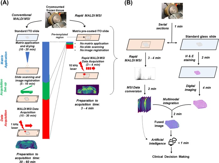
Matrix assisted laser desorption ionization mass spectrometry imaging (MALDI MSI) is an emerging analytical technique, which generates spatially resolved proteomic and metabolomic images from tissue specimens. Conventional MALDI MSI processing and data acquisition can take over 30 min, limiting its clinical utility for intraoperative diagnostics. We present a rapid MALDI MSI method, completed under 5 min, including sample preparation and analysis, providing a workflow compatible with the clinical frozen section procedure.

Keywords: Metabolomics; Molecular imaging; Molecular medicine; Surgical oncology.


Conflict of interest statement

Competing interestsD.S.C. and A.H. are employed at Bruker Daltonics. In compliance with Harvard Medical School and Partners Healthcare guidelines on potential conflict of interest, we disclose that N.Y.R.A. is a scientific advisor to BayesianDx and inviCRO. All other authors declare no competing interests.

Figures

Fig. 1
Fig. 1
a Comparison of workflow for conventional versus rapid MALDI MSI. The left panel illustrates additional steps and time needed for conventional MALDI MSI, including matrix application/drying, high-resolution slide scanning, image registration, and data acquisition using a 1 kHz laser. In the rapid MALDI MSI method (right panel), by using a matrix pre-coated slide and cryomounting the sample directly into a pre-templated region, and acquiring data with a 10 kHz laser, significant improvements in turnaround time are achieved. A representative ion image (for m/z 885.6) is illustrated below the workflow. Additional ion images comparing conventional and rapid method can be found in Supplemental Information. b Proposed workflow for rapid MALDI MSI in the frozen section room. Surgical specimens are serially cryosectioned onto a glass slide and a matrix pre-coated ITO slide. The section on the glass slide is stained using H&E followed by high-speed digital imaging, while the rapid MALDI MSI method is applied to the templated region on the ITO slide in a similar time frame. Following data acquisition, either pathologist-guided or artificial intelligence (AI)-guided diagnostic approaches may be utilized. In the pathologist-guided mode, the digital histological image is fused to the ion images using a rapid non-linear transformation (Supplemental Fig. 1), allowing the pathologist to select a region or pixel of interest for further molecular characterization. This approach could be used to quickly evaluate, for example, the presence or absence of cancer or of pathogens as well as particular molecular features of the cancer cells, a feat which is currently unachievable in a similar time frame. In the AI-guided mode (dashed arrow), the MSI data could be analyzed directly without visual review using previously machine trained models to classify regions based on their mass spectral signature
Fig. 2
Fig. 2
Histology and ion images generated by rapid MALDI MSI and t-distributed stochastic neighbor embedding (t-SNE) visualization of analyzed data. Coronal sections of a healthy mouse brain, and b mouse brain with a glioblastoma patient-derived xenograft. Surgically resected clinical research specimens of c normal breast tissue, d breast carcinoma, and e glioblastoma. The ion images were generated using SCiLs Lab software. Mass to charge ratio (m/z) of imaged analytes are listed on the left. Ions m/z 885.6 and m/z 888.7 represent PI(36:4) and ST(d42:2), respectively. Remaining ions are under investigation and will be identified using a combination of high-resolution mass spectrometry and fragmentation. Additional ion images are provided in the Supplemental Information, Suppl. Fig. 2

References

    1. Gal AA, Cagle PT. The 100-year anniversary of the description of the frozen section procedure. JAMA. 2005;294:3135. doi: 10.1001/jama.294.24.3135. - DOI - PubMed
    1. Brender, E. Frozen section biopsy. JAMA10.1001/jama.294.24.3200 (2005). - PubMed
    1. Yeh, Y. C. et al. Using frozen section to identify histological patterns in stage I lung adenocarcinoma of ≤3 cm: accuracy and interobserver agreement. Histopathology10.1111/his.12468 (2005). - PMC - PubMed
    1. St John, E. R. et al. Diagnostic accuracy of intraoperative techniques for margin assessment in breast cancer surgery a meta-analysis. Ann. Surg. 10.1097/SLA.0000000000001897 (2017). - PubMed
    1. Theisen, B. K., DiCianno, R. & Singhi, A. D. Pancreatic frozen section nightmares. Diagn. Histopathol.10.1016/j.mpdhp.2016.05.004 (2016).