. 2020 Mar 1;116(3):605-618.
doi: 10.1093/cvr/cvz182.

HAND1 loss-of-function within the embryonic myocardium reveals survivable congenital cardiac defects and adult heart failure

Affiliations

HAND1 loss-of-function within the embryonic myocardium reveals survivable congenital cardiac defects and adult heart failure

Beth A Firulli et al. Cardiovasc Res. .

Abstract

Aims: To examine the role of the basic Helix-loop-Helix (bHLH) transcription factor HAND1 in embryonic and adult myocardium.

Methods and results: Hand1 is expressed within the cardiomyocytes of the left ventricle (LV) and myocardial cuff between embryonic days (E) 9.5-13.5. Hand gene dosage plays an important role in ventricular morphology and the contribution of Hand1 to congenital heart defects requires further interrogation. Conditional ablation of Hand1 was carried out using either Nkx2.5 knockin Cre (Nkx2.5Cre) or α-myosin heavy chain Cre (αMhc-Cre) driver. Interrogation of transcriptome data via ingenuity pathway analysis reveals several gene regulatory pathways disrupted including translation and cardiac hypertrophy-related pathways. Embryo and adult hearts were subjected to histological, functional, and molecular analyses. Myocardial deletion of Hand1 results in morphological defects that include cardiac conduction system defects, survivable interventricular septal defects, and abnormal LV papillary muscles (PMs). Resulting Hand1 conditional mutants are born at Mendelian frequencies; but the morphological alterations acquired during cardiac development result in, the mice developing diastolic heart failure.

Conclusion: Collectively, these data reveal that HAND1 contributes to the morphogenic patterning and maturation of cardiomyocytes during embryogenesis and although survivable, indicates a role for Hand1 within the developing conduction system and PM development.

Keywords: HAND1; Transcription; Cardiac development; Heart failure with preserved ejection fraction; Mitral arcade; Right bundle branch block.


Figures

Figure 1
Figure 1
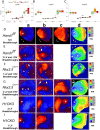
Cardiomyocyte-specific deletion of Hand1f/f and validation of select genes from transcriptome analysis of Nkx2.5Cre/+;Hand1f/f mutant hearts. (A) Wholemount in situ hybridization of control E10.5 embryo showing Hand1 LV expression (arrows). (B) Nkx2.5Cre; Hand1f/f E10.5 embryo shows a loss of visible Hand1 LV expression. (C) A similar Hand1f/f control as shown in A. (D) αMhc-Cre; Hand1f/f embryo shows a loss of visible Hand1 LV expression. (E) QRTPCR analysis was performed from E11.5 ventricle cDNA from Hand1f/f control and Nkx2.5Cre/+;Hand1f/f mutant hearts. Candidate expression analysis of genes associated with HAND1 regulation or not associated with an IPA biofunction category. (F) Expression analysis of genes chosen for protein kinase A and cardiac hypotrophy biofunctions. Error bars represent the minimum and maximum relative level of gene expression. N ≥ 6 (six mutants and six control hearts/primer set). Error bars denote the maximum and minimum relative level of gene expression in the test samples calculated using the confidence level set in the QuantStudio 3&5 software analysis settings. P values ≤0.05 generated by the QuantStudio 3 software which calculates Benjamini–Hochberg false discovery rate were regarded as significant and marked in all graphs as a single asterisk (*) n ≥ 6 in all experiments for all genotypes assayed. Scale bars = 150 μm.
Figure 2
Figure 2
Histological assessment of cardiac morphology reveals morphological defects in E14.5 H1CKOs hearts. H&E stained control E14.5 Hand1f/f hearts (A, n ≥ 10 and C, n ≥ 10) compared to Nkx2.5Cre; Hand1f/f (B, n ≥ 10); and αMhc-Cre; Hand1f/f (D, n ≥ 10) embryos. Control heart morphology shows distinct RV and LV and patent IVS. In Nkx2.5Cre; Hand1f/fmutants, ventricular septal defects are visible (asterisk), mitral valves appear immature (arrow), PM in B directly attaches to the valve (arrowhead). αMhc-Cre; Hand1f/f mutants (D) display similar ventricular septal defects (asterisks) and a PM directly inserting into the mitral valve (arrowhead n ≥ 10). Scale bars = 500 μm. Frequency of VSD occurrence was calculated to be 0.7 for Nkx2.5Cre generated and 0.5 for αMHC-Cre generated H1CKOs and exhibit significance with P values of 0.02 and 0.01, respectively obtained using the Fisher’s exact Test. (EH all n = 12 for controls n = 11 for mutants) immunohistochemistry sections of Control (E and F) and H1CKO (G and H) showing expression of the endothelial-specific protein CD31 (green) the cardiomyocyte-specific antibody to αMHC (MF20, red) and nuclei are visualized by DAPI staining (Blue). (I, n = 12 for control; n = 11 mutants). Sections were used to calculate the thickness of the LV and RV compact walls, RV and LV trabecular diameter, and the ratio of trabecula to compact wall. A Shapiro–Wilk test was applied to each set of measurements to test for normal distribution. All data sets fitting normal distribution were tested for statistical significance was calculated using unpaired t-test n = 12 for control and n = 11 mutant hearts. Measure of diameter of trabecular myocardium failed the normal distribution test, and significance was determined by Mann–Whitney U test. Scale bars = 500 and 50μm where indicated. Error bars represent standard deviation.
Figure 3
Figure 3
Functional analysis of Nkx2.5Cre; Hand1f/f mice show altered ventricular conduction. (A–C) Surface ECG analysis of leads I, II, and III from anaesthetized mice. QTc indicates corrected QT interval. A prolonged QRS duration is observed in Nkx2.5Cre; Hand1f/f mice (n = 6) in all leads (either QRSI or QRSII) when compared with controls (Hand1f/f n = 6; Nkx2.5Cre+/n = 5). A Shapiro–Wilk test was first applied to each set of measurements to test for normal distribution. Data were found to normal distribution and significance was determined using one-way ANOVA with post hoc Tukey HSD test. *P ≤ 0.05, **P ≤ 0.01. In the box-scatter plots centre lines indicate medians; box limits indicate the 25th and 75th percentiles; whiskers indicate the 1.5X the interquartile range from the box limits; dots show number of animals n ≥ 5. (D–I) Epicardial activation patterns in adult Hand1f/f(n = 6 each; D and E), Nkx2.5Cre+/ (n = 5 each; F and G) and Nkx2.5Cre; Hand1f/f hearts (n = 6 each; H and I) during atrial pacing. Shown are representative sequential optical maps (columns a–c) and the colour-coded activation maps (column d) for each genotype. LV and RV breakthroughs are demarked by asterisks and arrowheads, respectively. Optical maps were obtained during right atrial pacing at a cycle length of 120 ms or 150 ms. (D and E) One Hand1f/f heart showed a single breakthrough site on the anterior LV surface (D) consistent with the presence of a right bundle branch block, while another Hand1f/f heart showed two and one breakthrough sites on the left and right ventricular surface (E), respectively. (F and G) In Nkx2.5Cre+/ hearts, epicardial breakthrough occurred first on the right anterior surface (F) or manifested as double breakthrough sites on both the left and right ventricular anterior surfaces (G). (H and I) Three out of six Nkx2.5Cre; Hand1f/f (H1CKO) hearts showed two left ventricular breakthrough sites (H). The activation of the mid-anterior LV was grossly delayed (∼3 ms). The remaining three Nkx2.5Cre; Hand1f/f hearts exhibited single left ventricular breakthrough sites (I) indicative of right bundle branch block. Isochrone lines (I column d) demonstrate that LV global activation propagates from the LV to the RV. The prevalence of RBB was significantly higher in Nkx2.5Cre; Hand1f/f hearts compared with both Nkx2.5Cre and Hand1f/f hearts (P =0.002 by χ2 test followed by Fisher’s Exact tests for pairwise comparisons). Cross hairs present in some of the images are cursors and do not highlight any data. (D–I) Hand1f/f(n = 6; D and E), Nkx2.5Cre+/ (n = 5; F and G) and Nkx2.5Cre; Hand1f/f hearts (n = 6; H and I).
Figure 4
Figure 4
Adult (P60) Nkx2.5Cre; Hand1f/f mice exhibit cardiomegaly and mitral arcade. (A) Control Hand1f/f heart with unremarkable RA and LA and RV and LV. (B) Nkx2.5Cre; Hand1f/f heart is large and highly muscularized with a dilated LA, as well as RV and LV showing hypertrophic growth. (C) H&E section analysis of a control Hand1f/f heart shows normal chamber size and compaction. (D) Nkx2.5Cre; Hand1f/f heart displays thickened ventricular walls and both membranous (large arrowhead) and muscular (small arrowheads) VSDs. Boxed area in D is magnified in D’ showing PMs directly inserted into the mitral valve. (E) Control Hand1f/f lung showing normal morphology. (D) Nkx2.5Cre; Hand1f/f lung presents with marked pulmonary oedema. (G) A second Nkx2.5Cre; Hand1f/f example stained with Sirus red/fast green. VSD marked by arrowhead. Asterisks in G (magnified in H) show PM insertion into the mitral valve. (I) Magnified view of VSD shown in (G). (J) Echocardiography still image of a Nkx2.5Cre; Hand1f/f mutant show a large PM directly inserted into the MV (yellow arrowhead). Scale bars A–D and G, 650 μm; Scales bars E, F, H, and I, 100 μm. N ≥ 10.
Figure 5
Figure 5
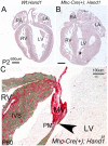
αMhc-Cre; Hand1f/f mice exhibit cardiomegaly and mitral arcade. (A) P2 H&E histology shows normal heart morphology. (B) αMhc-Cre; Hand1f/f mutant hearts display muscle overgrowth in the LV lumen and enlarged LA. (C) P60 Sirus red fast green stained αMhc-Cre; Hand1f/f mutant heart showing PM insertion into the MV and high levels of fibrosis in the IVS. Scale bars A and B 650 μm; Scales bars C 100 μm. N ≥ 10.
Figure 6
Figure 6
Documentation of cardiac defects living Nkx2.5Cre/+;Hand1f/f mutant hearts via cardiac ultrasound. (A) Still, image with schematic drawing to the right representing a patent cardiac outflow tract in Hand1f/f control heart. (B) Image from a similar location as shown in A of Nkx2.5Cre/+;Hand1f/f mutant that displays a membranous VSD (yellow arrowhead). (C) Still, image from a deeper plane showing a muscular VSD (yellow arrowhead) in a second Nkx2.5Cre/+;Hand1f/f mutant. (D) Pulse wave Doppler evaluation of a high-velocity blood jet from a membranous VSD measured from the mouse in Supplementary material online, Movie S1. (E) Example of EA reversal in a Nkx2.5Cre/+;Hand1f/f mutant. (F) M-mode evaluation of systolic function in a P ≥ 120 Nkx2.5Cre/+;Hand1f/f mutant showing poor systolic function with EF of only 38.79% and fractional shortening of 18.65%. Ao, aorta; LV, left ventricle; PA, pulmonary artery; RA, right atria; RV, right ventricle; TV, tricuspid valve (leaflets marked by asterisk); VSD, ventricular septal defect. N = 6.
Figure 7
Figure 7
Functional analysis of young Nkx2.5Cre; Hand1f/f mice show normal systolic function but altered peak aortic velocity. (A) Representative echocardiography still images of Hand1f/f control (n = 12, light grey), Nkx2.5Cre control (n = 12, open) and Nkx2.5Cre; Hand1f/f(n = 12, dark grey). (B) FS and EF at 4, 8, and 12 weeks of age show no changes in systolic function in Nkx2.5Cre; Hand1f/fcompared to controls. Doppler echocardiography measurement of aortic peak velocity (Ao peak) is significantly elevated (P = 0.04; one-way ANOVA) in mutants at 4 weeks and 12 weeks compared to controls. PA peak velocity is not significantly altered in Nkx2.5Cre; Hand1f/f mice. In the box-scatter plots centre lines indicate medians; box limits indicate the 25th and 75th percentiles; whiskers indicate the 1.5X the interquartile range from the box limits; dots show number of animals Hand1f/f control (n = 12, light grey), Nkx2.5Cre control (n = 12, open) and Nkx2.5Cre; Hand1f/f(n = 12, dark grey). (C) Tissue Doppler analysis of MV A at 4 weeks (asterisk) and MV E velocities at 4, 8, and 12 weeks are significantly elevated in Nkx2.5Cre; Hand1f/f mice compared to controls. Calculation of the mitral valve E/e’ ratio reveals that this measure of increased atrial filling pressure is also significantly higher in Nkx2.5Cre/+;Hand1f/f mice. *P ≤ 0.05 **P ≤ 0.01 via one-way ANOVA determined statistical significance with a P ≤ 0.05 confidence. A‘, peak velocity of diastolic mitral annular motion determined by pulse wave Doppler; E’, peak velocity of early diastolic mitral annular motion determined by pulse wave Doppler; E‘/A’, the E‘ to A’ ratio; IVRT, isovolumic relaxation time; MV A, peak velocity of late transmitral flow; MV Decel, deceleration time of early diastolic transmitral flow; MV E, peak velocity of early diastolic transmitral flow; MV E/A, ratio of MV E/A; MV E/E’, ratio of MV E/E'. ± indicates standard deviation. Hand1f/f control (n = 12, light grey), Nkx2.5Cre control (n = 12, open), and Nkx2.5Cre; Hand1f/f(n = 12, dark grey).

Comment in

References

    1. Olson EN. Gene regulatory networks in the evolution and development of the heart. Science 2006;313:1922–1927. - PMC - PubMed
    1. Pierpont ME, Basson CT, Benson DW Jr, Gelb BD, Giglia TM, Goldmuntz E, McGee G, Sable CA, Srivastava D, Webb CL; American Heart Association Congenital Cardiac Defects Committee; Council on Cardiovascular Disease in the Young. Genetic basis for congenital heart defects: current knowledge: a scientific statement from the American Heart Association Congenital Cardiac Defects Committee, Council on Cardiovascular Disease in the Young: endorsed by the American Academy of Pediatrics. Circulation 2007;115:3015–3038. - PubMed
    1. Gittenberger-de Groot AC, Blom NM, Aoyama N, Sucov H, Wenink AC, Poelmann RE.. The role of neural crest and epicardium-derived cells in conduction system formation. Novartis Found Symp 2008;250:125–134. discussion 134-141. - PubMed
    1. Scholl AM, Kirby ML.. Signals controlling neural crest contributions to the heart. Wires Syst Biol Med 2009;1:220–227. - PMC - PubMed
    1. Rochais F, Mesbah K, Kelly RG.. Signaling pathways controlling second heart field development. Circ Res 2009;104:933–942. - PubMed

Publication types

MeSH terms

Substances