

Cytotoxicity and proinflammatory effects of titanium and zirconia particles
- PMID: 31286286
- PMCID: PMC6614223
- DOI: 10.1186/s40729-019-0178-2
Cytotoxicity and proinflammatory effects of titanium and zirconia particles
Abstract
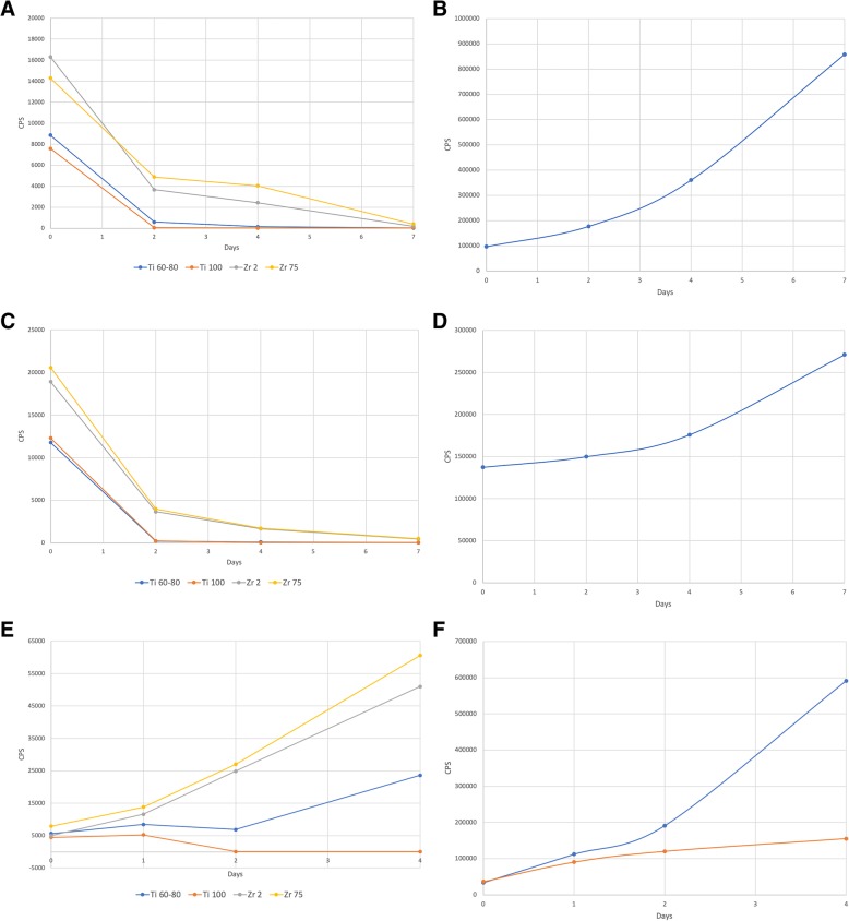
Background: To assess the effects of differently sized titanium (Ti) and zirconia (Zr) particles on (1) the metabolic activity of osteosarcoma-derived osteoblasts (SaOs-2) and human gingival fibroblasts (HGF) and (2) the cytokine expression of monocytes (THP-1) METHODS: Ti (60-80 nm and 100 nm) and Zr (2 μm and 75 μm) particles were incubated with SaOs-2, HGF, and THP-1 cells. At days 0, 2, 4, and 7 and 0, 1, 2, and 4 (THP-1), the mitochondrial activity was assessed and enzyme-linked immunosorbent assays were used to determine interleukin (IL)-1 beta and IL-6 concentrations of stimulated THP-1 at day 1.
Results: Ti60-80, Ti100, Zr2, and Zr75 particles were associated with gradual and significant within-group decreases in the viability of SaOs-2 and HGF cells. These effects were less pronounced in the Zr group. Similar to control cells, THP-1 did not reveal any significant increases in IL-1 beta and IL-6 concentrations. Viability of THP-1 was merely impaired in the presence of Ti100.
Conclusions: Ti and Zr particles had a detrimental effect on the viability of SaOs-2 and HGF, but no proinflammatory effect on THP-1.
Keywords: Cellular immunology; Cytokines; Fibroblast; In vitro model; Monocytes; Osteoblast.
Conflict of interest statement
Frank Schwarz, Maike Langer, Tina Hagena, Brigitte Hartig, Robert Sader, and Jürgen Becker declare that they have no competing interests related to this analysis.
Figures
References
LinkOut - more resources
Full Text Sources
