

Review
doi: 10.5152/dir.2019.18556.
Emerging clinical applications of high-intensity focused ultrasound
Affiliations
- PMID: 31287428
- PMCID: PMC6727814
- DOI: 10.5152/dir.2019.18556
Item in Clipboard
Review
Emerging clinical applications of high-intensity focused ultrasound
Diagn Interv Radiol.
2019 Sep.
Abstract
High-intensity focused ultrasound (HIFU) is a minimally-invasive and non-ionizing promising technology and has been assessed for its role in the treatment of not only primary tumors but also metastatic lesions under the guidance of ultrasound or magnetic resonance imaging. Its performance is notably effective in neurologic, genitourinary, hepato-pancreato-biliary, musculoskeletal, oncologic, and other miscellaneous applications. In this article, we reviewed the emerging technology of HIFU and its clinical applications.
Conflict of interest statement
The authors declared no conflicts of interest.
Figures
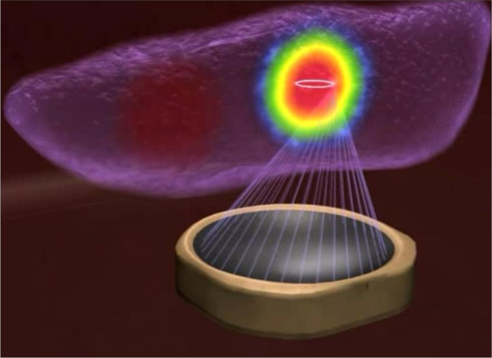
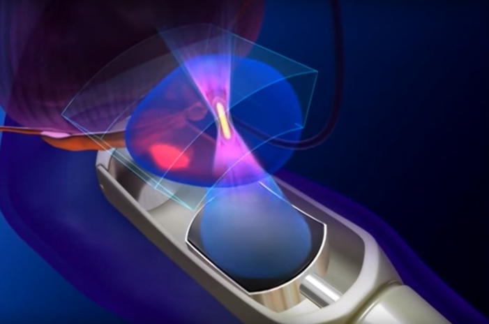
The transducer emits the therapeutic ultrasound beam to heat a focused point. (Figure courtesy of Profound Medical Corp – used with permission).
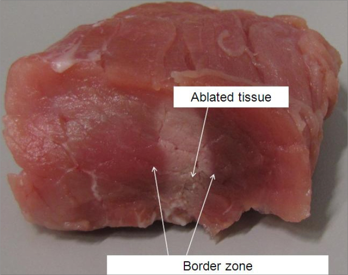
The immediate post-HIFU ablation image shows the HIFU-ablated region and the surrounding undamaged area. (Figure courtesy of Holger Grüll, University of Cologne – used with permission).
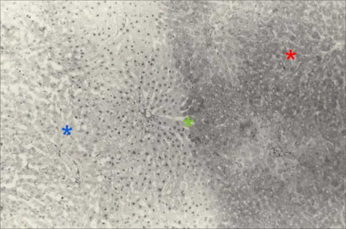
Histopathologic findings show the ablated region with irreversible cell damage comprised of deformed cells and pyknotic nuclei (blue asterisk), the borderline between the ablated and surrounding area with reversible cell damage (green asterisk), and the surrounding area with undamaged cells (red asterisk). (Figure courtesy of Lili Chen, Fox Chase Cancer Center – used with permission)
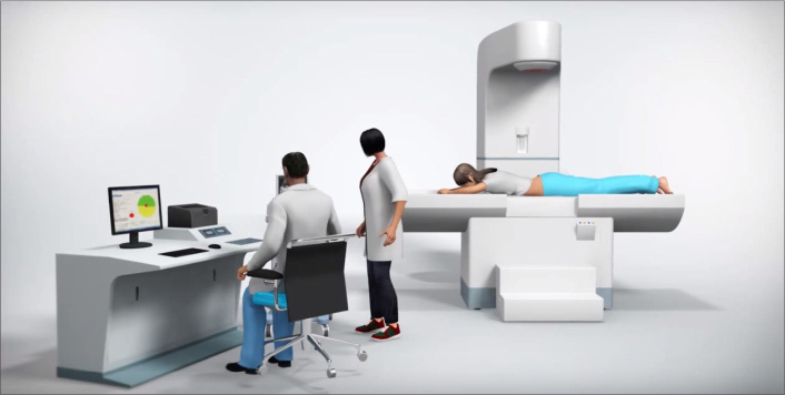
Ultrasound-guided HIFU system (Figure courtesy of Shenzhen Huikang Medical Apparatus Co., Ltd. – used with permission).
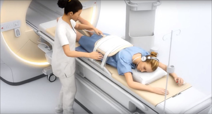
MRI-guided HIFU system (Figure courtesy of Profound Medical Corp – used with permission).
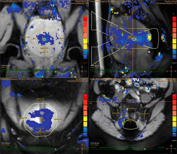
Real-time feedback temperature map of MRI-guided HIFU with the color scale during ablation therapy: upper left image shows coronal plane; upper right image shows sagittal plane; lower left image shows near field; lower right image shows far field.
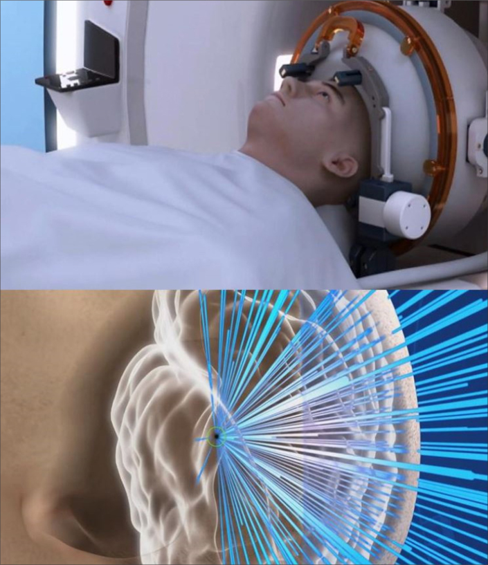
MRI-guided HIFU ablation for the brain with the ultrasound beam focused to the thalamus (Figure courtesy of Insightec Ltd. – used with permission).
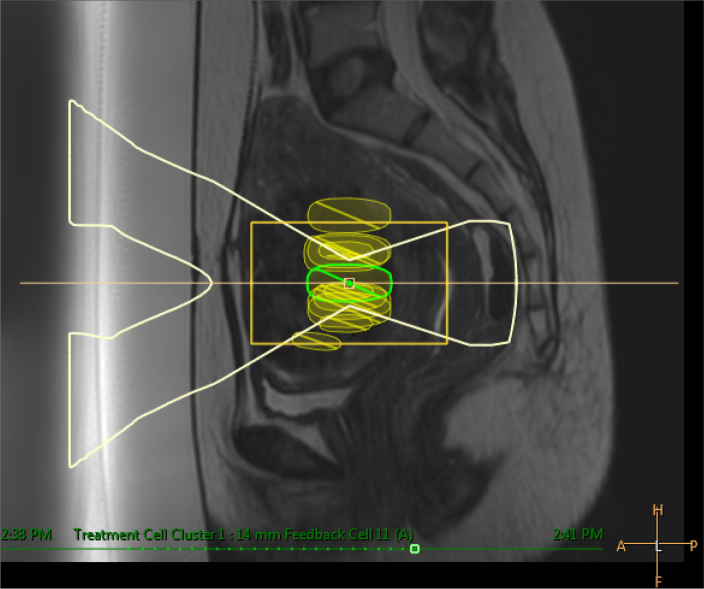
Sagittal T2-weighted image shows ultrasound beam under the guidance of MRI targeted to treatment cell located inside the uterine fibroid.
Transrectal ultrasound-guided HIFU ablation of the prostate with transrectal transducer (Figure courtesy of SonoCare Medical – used with permission).

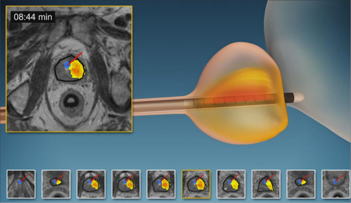
Transrectal MRI-guided HIFU ablation of the prostate with transrectal transducer along with temperature mapping during the ablation procedure (Figure courtesy of Insightec Ltd. – used with permission).
Transurethral MRI-guided HIFU ablation of the prostate with transurethral transducer along with temperature mapping during the ablation procedure (Figure courtesy of Profound Medical Corp – used with permission).
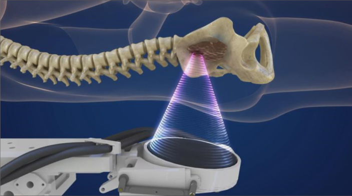
HIFU ablation of the bone with the ultrasound beam focused to the bone tumor so as to eradicate nerves inside the lesion (Figure courtesy of Insightec Ltd. – used with permission).

HIFU ablation of the breast tumor (Figure courtesy of Theraclion – used with permission).
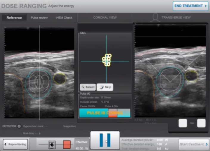
HIFU ablation of the thyroid nodule (Figure courtesy of Theraclion – used with permission).
References
-
- Wood RW, Loomis AL. The physical and biological effects of high frequency sound-waves of great intensity. Phil Mag. 1927;4:7–14. doi: 10.1080/14786440908564348. - DOI
Publication types
MeSH terms
LinkOut - more resources
Full Text Sources
