CO₂ Laser for the Treatment of Auricle Schwannoma: A Case Report and Review of the Literature
- PMID: 31287809
- PMCID: PMC6640171
- DOI: 10.12659/AJCR.916714
CO₂ Laser for the Treatment of Auricle Schwannoma: A Case Report and Review of the Literature
Abstract
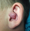
BACKGROUND Schwannoma, also called neuroma or neurolemmoma, is a tumor originating from the Schwann cells surrounding the nerves. It is an isolated benign tumor and its transformation into malignant cancer is very rare. Relatively uncommon, it is only the 5% of all the tumors of soft tissues. Its localization in the head and neck region accounts for up to 25-45% of schwannomas. In the outer ear, it commonly involves the external auditory canal, while auricle and tympanic membranes are very rare localizations of schwannomas. CASE REPORT We report a case of a 23-year-old male with a 3-year medical history of a growing neoplasm located in the left auricle concha, which was treated with a carbon dioxide laser (CO₂ laser) under local anesthesia. CONCLUSIONS Using a CO₂ laser allowed us to easily remove the tumor, reduce bleeding and surgical time, and avoid sutures and thus unsightly scars on the face. No complications and no relapse at 5 years of follow-up occurred.
Conflict of interest statement
None.
Figures


References
-
- Malone JP, Lee WJ, Levin RJ. Clinical characteristics and treatment outcome for nonvestibular schwannomas of the head and neck. Am J Otolaryngol. 2005;26:108–12. - PubMed
-
- Morais D, Santos J, Alonso M, et al. Schwannoma of the external auditory canal: An exceptional location. Acta Otorrinolaringol Esp. 2007;58(4):169–70. - PubMed
-
- Galli J, D’Ecclesia A, La Rocca LM, et al. Giant schwannoma of external auditory canal: A case report. Otolaryngol Head Neck Surg. 2001;124(4):473–74. - PubMed
-
- Biswas D, Marnane CN, Mal R, et al. Extracranial head and neck schwannomas: A 10-year review. Auris Nasus Larynx. 2007;34:353–59. - PubMed
-
- Yang CH, Su CY, Wei YC, et al. Schwannoma of the tympanic membrane. J Laryngol Otol. 2006;120(3):247–49. - PubMed
Publication types
MeSH terms
Substances
LinkOut - more resources
Full Text Sources

