. 2019 Jul 9;17(7):e3000337.
doi: 10.1371/journal.pbio.3000337. eCollection 2019 Jul.

Novel antibiotics effective against gram-positive and -negative multi-resistant bacteria with limited resistance

Affiliations

Novel antibiotics effective against gram-positive and -negative multi-resistant bacteria with limited resistance

Irène Nicolas et al. PLoS Biol. .

Abstract

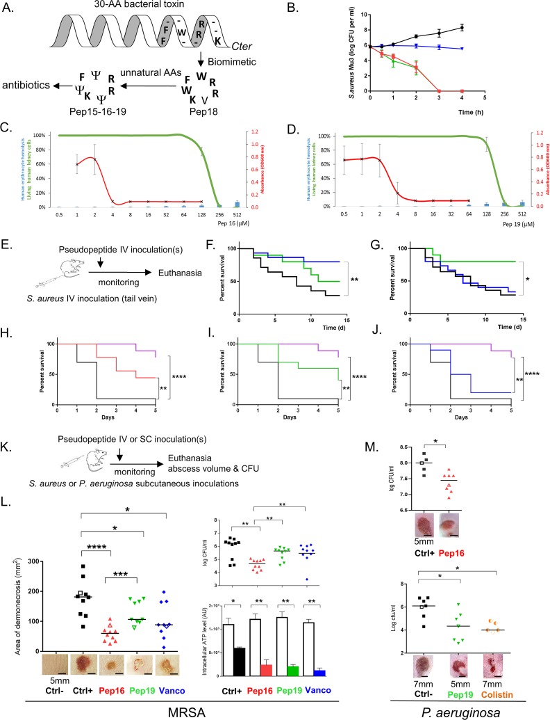
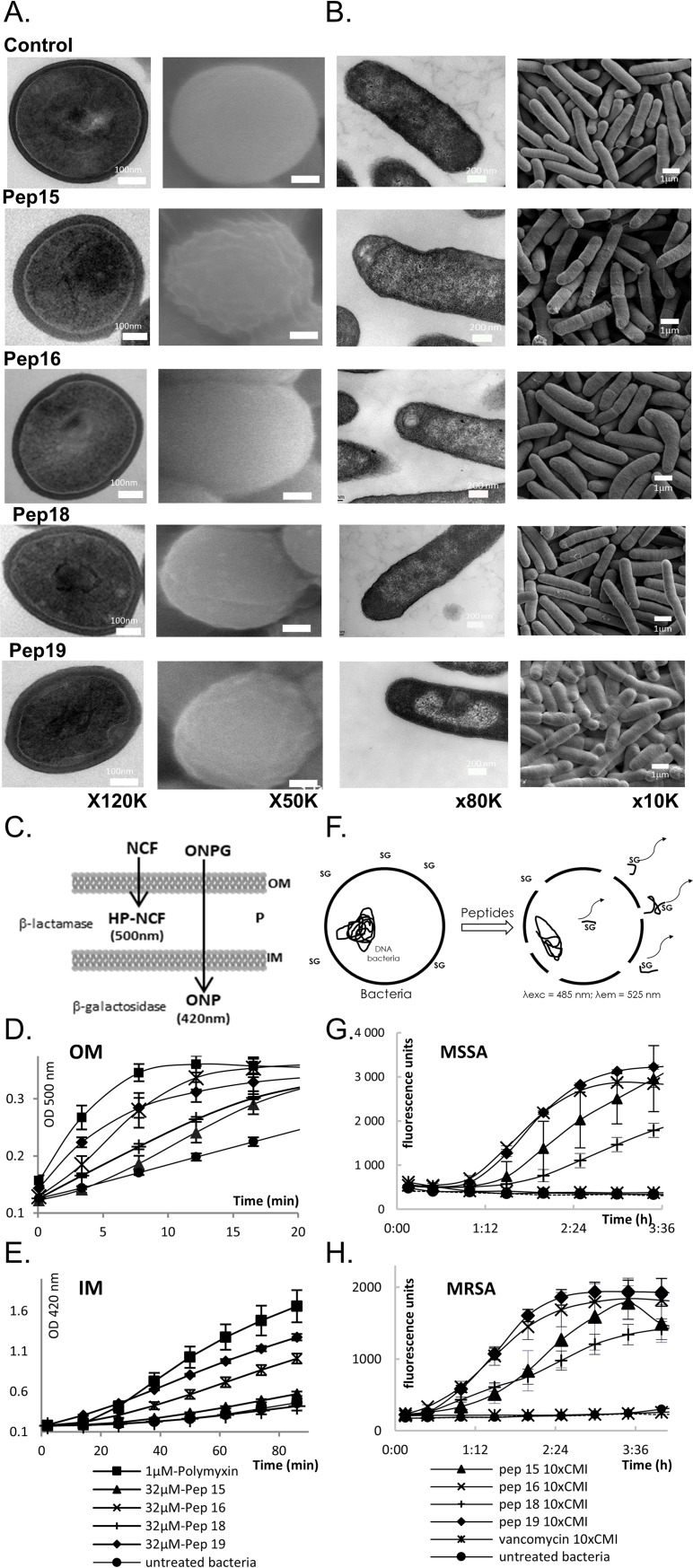
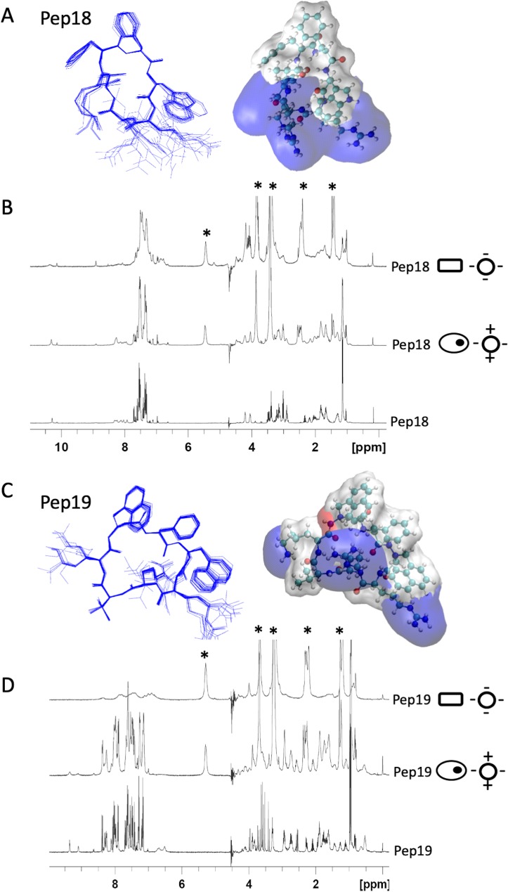
Antibiotics are a medical wonder, but an increasing frequency of resistance among most human pathogens is rendering them ineffective. If this trend continues, the consequences for public health and for the general community could be catastrophic. The current clinical pipeline, however, is very limited and is dominated by derivatives of established classes, the "me too" compounds. Here, we have exploited our recent identification of a bacterial toxin to transform it into antibiotics active on multidrug-resistant (MDR) gram-positive and -negative bacterial pathogens. We generated a new family of peptidomimetics-cyclic heptapseudopeptides-inspired from a natural bacterial peptide. Out of the 4 peptides studied, 2 are effective against methicillin-resistant Staphylococcus aureus (MRSA) in mild and severe sepsis mouse models without exhibiting toxicity on human erythrocytes and kidney cells, zebrafish embryos, and mice. These new compounds are safe at their active doses and above, without nephrotoxicity. Efficacy was also demonstrated against Pseudomonas aeruginosa and MRSA in a mouse skin infection model. Importantly, these compounds did not result in resistance after serial passages for 2 weeks and 4 or 6 days' exposure in mice. Activity of heptapseudopeptides was explained by the ability of unnatural amino acids to strengthen dynamic association with bacterial lipid bilayers and to induce membrane permeability, leading to bacterial death. Based on structure determination, we showed that cationic domains surrounded by an extended hydrophobic core could improve bactericidal activity. Because 2 peptide analogs, Pep 16 and Pep19, are effective against both MRSA and P. aeruginosa in severe sepsis and skin infection models, respectively, we believe that these peptidomimetics are promising lead candidates for drug development. We have identified potential therapeutic agents that can provide alternative treatments against antimicrobial resistance. Because the compounds are potential leads for therapeutic development, the next step is to start phase I clinical trials.


Conflict of interest statement

Cyclic peptides Pep15, Pep16, Pep18, and Pep19 are covered under European patent application #EP14191238.6, “Cyclic antimicrobial pseudopeptides and uses thereof.”

Figures

Fig 1
Fig 1. Two pseudopeptides are active against gram-positive and -negative MDR bacteria in murine sepsis and skin infection models and are nontoxic.
(A) Biomimetic design of antibiotics from a bacterial toxin [9]. Selected residues from the S. aureus toxin were cyclized, and unnatural amino acids (Ψ) were incorporated to synthesize those new antibiotics. Data associated with this panel can be found in S1 Fig. (B) Kill curves of MRSA using Pep16 (red), Pep19 (green), and vancomycin (blue) at concentrations 30-fold above the MIC are compared to untreated bacteria (black). Means and standard errors of the means are representative of 3 independent biological replicates. Data associated with this panel can be found in S3 Fig. (C–D) Pep16 and Pep19 antibiotic activity against MRSA strain Mu3 (red) compared to their low toxicity on human erythrocytes (blue bars) or to human kidney cell viability (green). Pep16 and Pep19 concentration range is from 0.5 to 512 μM. Data associated with this panel can be found in S4 Fig and S2–S6 Tables. (E) Overview of the mouse sepsis experiment. (F–G) Kaplan-Meier survival probability plots of mice infected with either MRSA without treatment (black), or with 1.5 mg.kg−1 Pep19 (green) or vancomycin (blue). In a sepsis protection model, groups of 5 Swiss mice were inoculated with approximately 5 × 10⁸ MRSA and treated either 3 h (F) or 15 h (G) post infection. Survival was monitored for 14 days after infection (panel d; x-axis), and the results are from 10 mice. The experiment was performed twice and the data combined. Data associated with these two panels can be found in S3 Fig. (H–J) Kaplan-Meier survival probability plots of severe sepsis assays (nearly all untreated infected mice killed within 2 days post infection) with mice infected with approximately 2.109 CFUs MRSA either without treatment (black) or with 4 repeated doses (0.5 mg.kg−1) of Pep16 (red), Pep19 (green), vancomycin (blue), or brilacidin (purple). The results are from 5 mice per assay, and the experiment was performed twice. Mice monitoring was performed for 5 days. (K) Overview of the mouse skin infection experiments with either S. aureus or P. aeruginosa. (L) Treatment of cutaneous abscesses from MRSA in mice using Pep16, Pep19, or vancomycin. Mice were infected with approximately 2 × 10⁹ MRSA and treated by IV 24 h post infection with a saline solution (black) or with 1.5 mg.kg−1 of Pep16 (red) or Pep19 (green) or vancomycin (blue). Lesion sizes and the CFU counts per abscess, plotted as individual points, were determined 6 days post infection (10 mice per condition). The abscess photos below the graph correspond to each experimental value shown in the graph as empty symbols. Intracellular ATP levels from S. aureus in the “normal-growing” (empty histograms) versus SCVs (filled colored histograms) collected from the mice skin abscesses (lower panels). (M) Treatment of cutaneous abscesses induced by P. aeruginosa in mice using Pep16, Pep19, or colistin. Mice were infected with approximately 108 P. aeruginosa and treated with repeated doses post infection with a saline solution (black) or with Pep16 (red, 30 mg.kg−1), Pep19 (green, 30 mg.kg−1), or colistin (orange, 9 mg.kg−1, lower panel). The CFU counts per abscess, plotted as individual points, were determined 3 days post infection (5 to 8 mice per condition). The abscess photos below the graph correspond to each experimental value shown as empty symbols. Mann-Whitney was used to calculate the differences between the groups; *0.05 < P < 0.01; **0.01 < P < 0.001; ***0.001 < P < 0.0001; ****P < 0.00001. Data associated with this figure can be found in S1 Data. CFU, colony-forming unit; Ctrl, controls; MDR, multidrug resistant; MIC, minimal inhibitory concentration; MRSA, methicillin-resistant S. aureus; OD600, optical density at 600 nm; SCV, small colony variant.
Fig 2
Fig 2. Pseudopeptide actions on gram-negative and -positive bacteria.
Cell wall and membrane alterations of S. aureus Newman (A) and E. coli K12 (B), suggested by typical and representative TEM (left) and SEM (right) micrographs after treatment for 2 h at 37°C with Pep15, Pep16, Pep18, or Pep19 at their MICs. Untreated bacteria are unaltered, and cell walls and membranes remain intact. Pep15 and Pep19 erode cell walls (black arrows), and Pep19 triggers intracellular perturbations (white arrow). Data associated with panel (A) can be found in S6 Fig. Overview panels of the leakage assays for E. coli (C) and S. aureus (F). (C–H) Peptidomimetics permeate gram-negative (E. coli) OMs and IMs (panels C–E) and gram-positive MSSA and MRSA membranes (panels F–H). (C) Overview of the leakage experiments on gram-negative bacteria. (D) NCF degradation products triggered by periplasmic β-lactamases. (E) ONPG degradation products produced by cytoplasmic β-galactosidases. The mean values of 3 independent experiments are presented. Shown are polymyxin B as positive control, untreated bacteria, and Pep15, Pep16, Pep18, or Pep19. (F) Overview of the leakage experiments on gram-positive MSSA and MRSA. SG cannot enter intact cells but bind DNA and fluoresce once the cell integrity is damaged. MSSA (panel G) and MRSA (panel H) leakage experiments evidencing pseudopeptide-induced membrane alterations. On both gram-negative and -positive bacteria, the highest activity is with Pep16 and Pep19. Data associated with this figure can be found in S1 Data. IM, inner membrane; MIC, minimal inhibitory concentration; MRSA, methicillin-resistant S. aureus; MSSA, methicillin-susceptible S. aureus; NCF, nitrocefin; OM, outer membrane; ONPG, O-nitrophenyl-β-D-galactopyranoside; SEM, scanning electron microscopy; SG, sytox green; TEM, transmission electron microscopy.
Fig 3
Fig 3. Three-dimensional structures of two pseudopeptides and their dynamics with eukaryotic and prokaryotic membrane mimics.
NMR structures of Pep18 (A) and Pep19 (C) analogs in presence of SDS micelles. These differ only in the replacement of 2 phenylalanine residues in Pep18 by 2 aza-β3-1-naphthylalanyl residues in Pep19. Pep18 cyclic backbone has 7 natural amino acids. Two hydrogen bonds involve Val6 carbonyl oxygen with HN Phe2 (residue i + 2) and HN Trp3 (residue i + 3). Two aza-β3-naphthylalanine residues from Pep19 form 2 R-hydrazino turns with 2 hydrogen bonds between the amide proton of residue i with the lone pair of the sp3 nitrogen atom of residue (i − 1) and the carbonyl of residue (i − 2). One of the hydrazino turns involves residues 2–4 and is reinforced by an additional bond between the carbonyl of Phe2 and the amine of Arg5. The ternary nitrogen configuration of the aza-β3-amino acids is an R absolute configuration. The electrostatic potential surface maps of Pep18 and Pep19 in the presence of SDS micelles are depicted with blue cationic areas and white hydrophobic domains. (B, D) 1H NMR spectra of 2 mM peptide analogs (panel B: Pep18; panel D: Pep19) without SUV (lower spectra), in the presence of zwitterionic SUVs (middle; 8 mM DMPC-d54) or anionic SUVs (top; 8 mM DMPC-d54/DMPS-d54 70:30). Zwitterionic and anionic SUVs mimic eukaryotic (ovals with an inner black dot) and bacterial (rectangles) membranes, respectively. “*” corresponds to the lipid signals. Data associated with this figure can be found in S8–S11 Tables. Arg,arginine 5; DMPC-d54, 1,2-dimyristoyl-d54-sn-glycero-3-phosphocholine; DMPS-d54, 1,2-dimyristoyl-d54-sn-glycero-3-phosphoserine; HP-NCF, hydrolyzed product–nicrocefin; NMR, nuclear magnetic resonance; Phe2, phenylalanine 2; ppm, parts per million; SDS, sodium dodecyl sulfate; SUV, small unilamellar vesicle.

Similar articles

Cited by

References

    1. Levy SB, Marshall B. Antibacterial resistance worldwide: causes, challenges and responses. Nat Med. 2004;10(12 Suppl):S122–9 10.1038/nm1145 - DOI - PubMed
    1. Blair JM, Webber MA, Baylay AJ, Ogbolu DO, Piddock LJ. Molecular mechanisms of antibiotic resistance. Nat Rev Microbiol. 2015;13(1):42–51 10.1038/nrmicro3380 - DOI - PubMed
    1. Boucher HW, Talbot GH, Bradley JS, Edwards JE, Gilbert D, Rice LB, et al. Bad bugs, no drugs: no ESKAPE! An update from the Infectious Diseases Society of America. Clin Infect Dis. 2009;48(1):1–12. 10.1086/595011 - DOI - PubMed
    1. Khan SN, Khan AU. Breaking the Spell: Combating Multidrug Resistant 'Superbugs'. Front Microbiol. 2016;7:174 10.3389/fmicb.2016.00174 - DOI - PMC - PubMed
    1. de Kraker ME, Stewardson AJ, Harbarth S. Will 10 Million People Die a Year due to Antimicrobial Resistance by 2050? PLoS Med. 2016;13(11):e1002184 10.1371/journal.pmed.1002184 - DOI - PMC - PubMed

Publication types

MeSH terms

Substances