. 2019 Jul 9;14(7):e0219572.
doi: 10.1371/journal.pone.0219572. eCollection 2019.

PKCζ and JNK signaling regulate radiation-induced compensatory proliferation in parotid salivary glands

Affiliations

PKCζ and JNK signaling regulate radiation-induced compensatory proliferation in parotid salivary glands

Wen Yu Wong et al. PLoS One. .

Abstract

Radiotherapy is a common treatment option for head and neck cancer patients; however, the surrounding healthy salivary glands are often incidentally irradiated during the process. As a result, patients often experience persistent xerostomia and hyposalivation, which deceases their quality of life. Clinically, there is currently no standard of care available to restore salivary function. Repair of epithelial wounds involves cellular proliferation and establishment of polarity in order to regenerate the tissue. This process is partially mediated by protein kinase C zeta (PKCζ), an apical polarity regulator; however, its role following radiation damage is not completely understood. Using an in vivo radiation model, we show a significant decrease in active PKCζ in irradiated murine parotid glands, which correlates with increased proliferation that is sustained through 30 days post-irradiation. Additionally, salivary glands in PKCζ null mice show increased basal proliferation which radiation treatment did not further potentiate. Radiation damage also activates Jun N-terminal kinase (JNK), a proliferation-inducing mitogen-activated protein kinase normally inhibited by PKCζ. In both a PKCζ null mouse model and in primary salivary gland cell cultures treated with a PKCζ inhibitor, there was increased JNK activity and production of downstream proliferative transcripts. Collectively, these findings provide a potential molecular link by which PKCζ suppression following radiation damage promotes JNK activation and radiation-induced compensatory proliferation in the salivary gland.


Conflict of interest statement

The authors have declared that no competing interests exist.

Figures
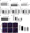
Fig 1
Fig 1. Radiation decreases pPKCζ-T560 but not total levels of the PKCζ/Par3/Par6 complex.
FVB mice were either untreated (UT) or irradiated (IR) with 5Gy and dissected on days 4, 5, 7, and 30 following radiation treatment. Total protein levels of (A-B) Par3, (C-D) Par6, (E-F), total PKCζ, and (G-H) pPKCζ-T560 were evaluated following radiation treatment. Immunoblots were re-probed with β-tubulin or PKCζ as a loading control. (I) Immunofluorescent staining was used to determine the intensity area of pPKCζ-T560 (red) in comparison to the total area. Composite images with DAPI (blue) are presented in both high and low magnification views (scale bar for high magnification = 30 μm, low magnification = 100 μm). (J) Quantification of the percentage of pPKCζ-T560 positive area within the parotid gland. Results are presented from at least three mice per condition; I-J used 10–15 images per mouse; error bars denote mean ± SEM. Significant difference (p<0.05) was determined by a Tukey-Kramer test for multiple comparisons. Treatment groups with different letters above the bar graphs are significantly different from each other.
Fig 2
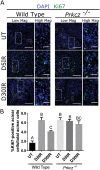
Fig 2. Depletion of PKCζ induces proliferation in vivo.
Wild Type C57BL/6J and Prkcz -/- mice were either untreated (UT) or irradiated (IR) with 5Gy and dissected on days 5 and 30 following radiation treatment. (A) Immunofluorescent staining with Ki67 (green) was used to determine the number of proliferating cells in comparison to the total number of cells in the acinar compartment. Composite images with DAPI (blue) are presented in both high and low magnification views (scale bar for high magnification = 30 μm, low magnification = 100 μm). The yellow dotted outline represents the ductal compartment while the rest of the glandular area represents the acinar compartment. (B) Quantification of A. Results are presented from at least four mice per condition; A-B used 10–15 images per mouse; error bars denote mean ± SEM. Significant difference (p<0.05) was determined by a Tukey-Kramer test for multiple comparisons. Treatment groups with different letters above the bar graphs are significantly different from each other.
Fig 3
Fig 3. Radiation induces JNK signaling in vivo.
FVB mice were either untreated (UT) or irradiated (IR) with 5Gy and dissected on day 5 following radiation treatment to evaluate JNK signaling. (A) Relative JNK kinase activity was measured by incubating immunoprecipitated JNK in the presence of ATP and a c-Jun substrate. The amount of phosphorylated c-Jun (S73) was detected via immunoblots. (B) Levels of phosphorylated c-Jun (S63) were determined following radiation treatment. Total c-Jun was probed as a loading control. (C) Relative mRNA levels of MT1F, PDE3A, NFATC2, and CCND1 were determined by RT-PCR and normalized to GAPDH. Results are presented from at least four mice per condition. Significant difference (p<0.05) was determined by Student’s t-test. *(p<0.05), **(p<0.01).
Fig 4
Fig 4. Inhibition of JNK signaling with SP600125 in irradiated cells.
Parotid salivary glands from FVB mice were dissected and cultured as primary cell cultures. One day after dissection, the primary cells were irradiated with 5Gy and cell lysates or RNA were collected on Day 5 after radiation treatment. On Day 4 after radiation treatment, the cells were either treated with 10 μM SP600125 or DMSO vehicle control. (A) Effects of SP600125 treatment on phosphorylated c-Jun (S63) in primary salivary cells were evaluated by immunoblotting. Blots were reprobed for total levels of c-Jun as a loading control. (B) Quantification by densitometry of A normalized to DMSO vehicle control. (C-F) Relative MT1F, PDE3A, NFATC2, and CCND1 mRNA levels determined by RT-PCR and normalized to GAPDH. Results are presented from at least three independent primary cell preparations; error bars denote mean ± SEM. Significant difference (<0.05) was determined by a Tukey-Kramer test for multiple comparisons. Treatment groups with different letters above the bar graphs are significantly different from each other.
Fig 5
Fig 5. Modulation of PKCζ increases JNK signaling.
Parotid salivary glands from FVB mice were dissected and cultured as primary cell cultures. At sub-confluency, the cells were treated with 20 μM PKCζ pseudosubstrate inhibitor (PPI) or vehicle control. (A) Relative JNK kinase activity was measured by incubating immunoprecipitated JNK in the presence of ATP and c-Jun substrate in cells treated with 20 μM PPI or vehicle control. The amount of phosphorylated c-Jun (S73) was detected via immunoblots. (B) Total protein levels for phosphorylated c-Jun (S63) was determined following PPI inhibition. Immunoblots were reprobed with total c-Jun as a loading control. Relative quantification is depicted below the blot. (C) Relative mRNA levels of MT1F, PDE3A, NFATC2, and CCND1 were determined by RT-PCR and normalized to GAPDH as a loading control. Lysates and mRNA were collected from C57BL/6J wildtype and Prkcz -/- mice. (D) Relative JNK kinase activity was measured by incubating immunoprecipitated JNK in the presence of ATP and c-Jun substrate in wildtype or Prkcz -/- mice. The amount of phosphorylated c-Jun (S73) was detected via immunoblots. (E) Total protein levels for phosphorylated c-Jun (S63) was determined in wildtype or Prkcz -/- mice. Immunoblots were reprobed with total c-Jun as a loading control. Relative quantification is depicted below the blot. (F) Relative mRNA levels of MT1F, PDE3A, NFATC2, and CCND1 were determined by RT-PCR and normalized to GAPDH as a loading control. Results are presented from at least four mice for in vivo experiments or three independent in vitro primary cell culture experiments per condition; error bars denote mean ± SEM. Significant difference (p<0.05) was determined by Student’s t-test. *(p<0.05), **(p<0.01).

Similar articles

Cited by

References

    1. Lo Nigro C, Denaro N, Merlotti A, Merlano M. Head and neck cancer: Improving outcomes with a multidisciplinary approach. Cancer Manag Res. 2017;9: 363–371. 10.2147/CMAR.S115761 - DOI - PMC - PubMed
    1. Denaro N, Merlano MC, Russi EG. Follow-up in head and neck cancer: Do more does it mean do better? A systematic review and our proposal based on our experience. Clin Exp Otorhinolaryngol. 2016;9: 287–297. 10.21053/ceo.2015.00976 - DOI - PMC - PubMed
    1. Belcher R, Hayes K, Fedewa S, Chen AY. Current treatment of head and neck squamous cell cancer. J Surg Oncol. 2014;110: 551–574. 10.1002/jso.23724 - DOI - PubMed
    1. Lin H, Limesand K, Ann D. Current State of Knowledge on Salivary Gland Cancers. Crit Rev Oncog. 2018;23: 139–151. 10.1615/CritRevOncog.2018027598 - DOI - PMC - PubMed
    1. Haynie JL, Bryant PJ. The effects of X-rays on the proliferation dynamics of cells in the imaginal wing disc of Drosophila melanogaster. Wilhelm Roux’s Arch Dev Biol. 1977;183: 85–100. 10.1007/BF00848779 - DOI - PubMed

Publication types

MeSH terms