. 2019 Jul 8;29(13):2199-2207.e10.
doi: 10.1016/j.cub.2019.05.058.

Reconstitution of Microtubule Nucleation In Vitro Reveals Novel Roles for Mzt1

Affiliations

Reconstitution of Microtubule Nucleation In Vitro Reveals Novel Roles for Mzt1

Su Ling Leong et al. Curr Biol. .

Erratum in

Abstract

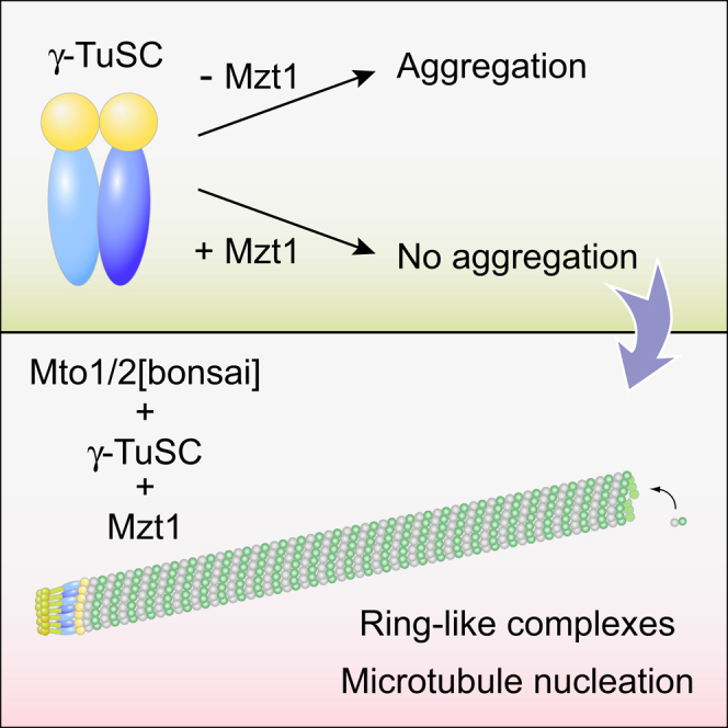
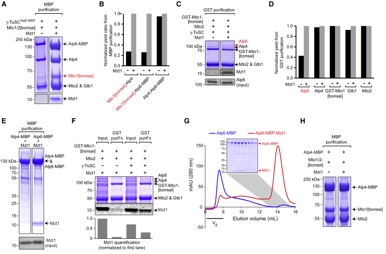
Microtubule (MT) nucleation depends on the γ-tubulin complex (γ-TuC), in which multiple copies of the heterotetrameric γ-tubulin small complex (γ-TuSC) associate to form a ring-like structure (in metazoans, γ-tubulin ring complex; γ-TuRC) [1-7]. Additional conserved regulators of the γ-TuC include the small protein Mzt1 (MOZART1 in human; GIP1/1B and GIP2/1A in plants) [8-13] and proteins containing a Centrosomin Motif 1 (CM1) domain [10, 14-19]. Many insights into γ-TuC regulators have come from in vivo analysis in fission yeast Schizosaccharomyces pombe. The S. pombe CM1 protein Mto1 recruits the γ-TuC to microtubule-organizing centers (MTOCs) [14, 20-22], and analysis of Mto1[bonsai], a truncated version of Mto1 that cannot localize to MTOCs, has shown that Mto1 also has a role in γ-TuC activation [23]. S. pombe Mzt1 interacts with γ-TuSC and is essential for γ-TuC function and localization to MTOCs [11, 12]. However, the mechanisms by which Mzt1 functions remain unclear. Here we describe reconstitution of MT nucleation using purified recombinant Mto1[bonsai], the Mto1 partner protein Mto2, γ-TuSC, and Mzt1. Multiple copies of the six proteins involved coassemble to form a 34-40S ring-like "MGM" holocomplex that is a potent MT nucleator in vitro. Using purified MGM and subcomplexes, we investigate the role of Mzt1 in MT nucleation. Our results suggest that Mzt1 is critical to stabilize Alp6, the S. pombe homolog of human γ-TuSC protein GCP3, in an "interaction-competent" form within the γ-TuSC. This is essential for MGM to become a functional nucleator.


Figures

None
Graphical abstract
Figure 1
Figure 1
Mzt1 Prevents Large-Scale Aggregation of the γ-TuSC In Vitro (A) SDS-PAGE of γ-TuSCAlp6-MBP (without Mzt1), purified on amylose via Alp6-MBP. (B) Superose 6 size-exclusion chromatography (SEC) of γ-TuSCAlp6-MBP, with corresponding SDS-PAGE of indicated fractions. The majority of eluting γ-TuSCAlp6-MBP elutes in the void volume. (C) SDS-PAGE of 80-min density-gradient centrifugation of γ-TuSCAlp6-MBP (SYPRO Ruby stain). Most γ-TuSC is in the pellet. Results from a 45-min centrifugation are shown in Figure S1C. (D) SDS-PAGE of γ-TuSCAlp6-MBP with Mzt1 (γ-TuSCAlp6-MBP:Mzt1), purified as above. Mzt1 is shown from a higher-contrast image, shown in Figure S1B. (E) Superose 6 SEC of (γ-TuSCAlp6-MBP:Mzt1), with corresponding SDS-PAGE of indicated fractions. Alp6-MBP, Alp4, γ-tubulin (Gtb1), and Mzt1 coelute, much later than the void volume. (F) SDS-PAGE of 80-min density-gradient centrifugation of γ-TuSCAlp6-MBP:Mzt1 (SYPRO Ruby stain). γ-TuSCAlp6-MBP:Mzt1 sediments with a broad profile centered at ∼24S. Mzt1 was visualized on a separate gel with higher acrylamide concentration. Results from a 45-min centrifugation are shown in Figure S1D. See also Figure S1.
Figure 2
Figure 2
γ-TuSC:Mzt1 Interacts with Mto1/2[bonsai] Complex to Form An “MGM” (Mto/Gamma/Mzt) Holocomplex (A) SDS-PAGE of 80-min density-gradient centrifugation of Mto1/2[bonsai]. (B) SDS-PAGE of 80-min density-gradient centrifugation of Mto1/2[bonsai] mixed (after purification) with γ-TuSCAlp6-MBP:Mzt1. Mixing alters the sedimentation profiles of both complexes, and all constituent proteins cosediment. Mzt1 was visualized on a separate gel with higher acrylamide concentration. Gels are stained with SYPRO Ruby. Representative sedimentation profiles from (A) and (B) are shown in Figure S2A. (C) Interactions between different proteins within MGM holocomplex, identified by zero-length chemical crosslinking and mass spectrometry (see STAR Methods). For simplicity, only interprotein crosslinks are shown. In this experiment, crosslinks to Mzt1 (a very small protein with few useful proteolytic fragments) were not identified. (D) Negative-stain electron microscopy of MGM. The image is a portion of a field shown in Figure S2F. In (C) and (D), MGM was purified from coexpression of Mto1/2[bonsai] and γ-TuSCAlp6-MBP:Mzt1 proteins. See also Figures 1 and S2.
Figure 3
Figure 3
The MGM Holocomplex Is a Potent Microtubule Nucleator In Vitro (A) DAPI fluorescence assay for microtubule (MT) polymerization, using porcine brain tubulin and the indicated complexes and concentrations. The MGM holocomplex nucleates MTs in a dose-dependent manner. The estimated molarity of MGM is based on assumption that Mto1/2[bonsai] and γ-TuSCAlp6-MBP:Mzt1 coassemble into a structure similar to the mammalian γ-TuRC (see STAR Methods). Additional controls are shown in Figures S3A–S3C. (B) Rhodamine-tubulin fluorescence microscopy assay for MT polymerization. (C) Quantification of MT polymerization for experiments in (B). Each data point represents total MT fluorescence within a randomly chosen field. Inset shows expanded scale for non-MGM samples. Quantification of MT number per field is shown in Figure S3D. Scale bar, 10 μm. See also Figure S3.
Figure 4
Figure 4
Mzt1 Stabilizes Alp6 in an “Interaction-Competent” State within Higher-Order Complexes (A) SDS-PAGE of amylose (MBP) purification of coexpressed γ-TuSCAlp6-MBP and Mto1/2[bonsai] in the absence versus presence of Mzt1. Mzt is required for efficient copurification of Mto1[bonsai] with γ-TuSC. Note that the total yield of γ-TuSC proteins is also decreased. Western blots of protein inputs are shown in Figure S4A. (B) Quantification of copurification of the indicated proteins from (A). Values for copurification are expressed as ratios, to correct for differences in yield, and normalized to values obtained in the presence of Mzt1. (C) SDS-PAGE and anti-Mzt1 immunoblot of glutathione-agarose (GST) purification of coexpressed Mto1/2[bonsai]GST-Mto1[bonsai] and γ-TuSC in the absence versus presence of Mzt1. Mzt is required for efficient copurification of Alp6 with Mto1/2[bonsai]. Anti-Alp6 western blot indicates comparable Alp6 input levels in absence versus presence of Mzt1. (D) Quantification of copurification of the indicated proteins in (C). Quantification was based on western blots shown in Figure S4B. Equivalent experiments using Mto1/2[bonsai]Strep-Mto1[bonsai] are shown in Figures S4C–S4E. (E) SDS-PAGE of amylose (MBP) purification of Alp4-MBP or Alp6-MBP coexpressed with Mzt1. Mzt1 copurifies with Alp6-MBP but not with Alp4-MBP. Anti-Mzt1 western blot indicates comparable Mzt1 levels in the two inputs. (F) SDS-PAGE and anti-Mzt1 western blot of glutathione-agarose purification of coexpressed Mto1/2[bonsai]GST-Mto1[bonsai] and Mzt1 in the absence versus presence of γ-TuSC. Mzt1 does not copurify with Mto1/2[bonsai] unless γ-TuSC is also present. The graph shows quantification of anti-Mzt1 western blot, normalized to value in first lane. (G) Superose 6 size-exclusion chromatography of Alp6-MBP purified alone (blue) or from coexpression with Mzt1 (red). Alp6-MBP elutes in void volume, while Alp6-MBP:Mzt1 elutes much later. Inset shows SDS-PAGE of indicated fractions for Alp6-MBP:Mzt1. (H) SDS-PAGE of amylose (MBP) purification of coexpressed Alp4-MBP and Mto1/2[bonsai] in the absence versus presence of Mzt1. Mto1/2[bonsai] copurifies with Alp4-MBP, independent of Mzt1 and other γ-TuSC proteins. The control experiment for non-specific binding of Mto1/2[bonsai] to amylose is shown in Figure S4H. See also Figure S4.

Similar articles

Cited by

References

    1. Kollman J.M., Merdes A., Mourey L., Agard D.A. Microtubule nucleation by γ-tubulin complexes. Nat. Rev. Mol. Cell Biol. 2011;12:709–721. - PMC - PubMed
    2. Kollman, J.M., Merdes, A., Mourey, L., and Agard, D.A. (2011). Microtubule nucleation by γ-tubulin complexes. Nat. Rev. Mol. Cell Biol. 12, 709-721. - PMC - PubMed
    1. Petry S., Vale R.D. Microtubule nucleation at the centrosome and beyond. Nat. Cell Biol. 2015;17:1089–1093. - PubMed
    2. Petry, S., and Vale, R.D. (2015). Microtubule nucleation at the centrosome and beyond. Nat. Cell Biol. 17, 1089-1093. - PubMed
    1. Lin T.C., Neuner A., Schiebel E. Targeting of γ-tubulin complexes to microtubule organizing centers: conservation and divergence. Trends Cell Biol. 2015;25:296–307. - PubMed
    2. Lin, T.C., Neuner, A., and Schiebel, E. (2015). Targeting of γ-tubulin complexes to microtubule organizing centers: conservation and divergence. Trends Cell Biol. 25, 296-307. - PubMed
    1. Roostalu J., Surrey T. Microtubule nucleation: beyond the template. Nat. Rev. Mol. Cell Biol. 2017;18:702–710. - PubMed
    2. Roostalu, J., and Surrey, T. (2017). Microtubule nucleation: beyond the template. Nat. Rev. Mol. Cell Biol. 18, 702-710. - PubMed
    1. Farache D., Emorine L., Haren L., Merdes A. Assembly and regulation of γ-tubulin complexes. Open Biol. 2018;8:170266. - PMC - PubMed
    2. Farache, D., Emorine, L., Haren, L., and Merdes, A. (2018). Assembly and regulation of γ-tubulin complexes. Open Biol. 8, 170266. - PMC - PubMed

Publication types

MeSH terms