Review
. 2019 Jul 8;29(13):R647-R662.
doi: 10.1016/j.cub.2019.05.026.

Evolution of the Chordate Telencephalon

Affiliations
Review

Evolution of the Chordate Telencephalon

Steven D Briscoe et al. Curr Biol. .

Abstract

The dramatic evolutionary expansion of the neocortex, together with a proliferation of specialized cortical areas, is believed to underlie the emergence of human cognitive abilities. In a broader phylogenetic context, however, neocortex evolution in mammals, including humans, is remarkably conservative, characterized largely by size variations on a shared six-layered neuronal architecture. By contrast, the telencephalon in non-mammalian vertebrates, including reptiles, amphibians, bony and cartilaginous fishes, and cyclostomes, features a great variety of very different tissue structures. Our understanding of the evolutionary relationships of these telencephalic structures, especially those of basally branching vertebrates and invertebrate chordates, remains fragmentary and is impeded by conceptual obstacles. To make sense of highly divergent anatomies requires a hierarchical view of biological organization, one that permits the recognition of homologies at multiple levels beyond neuroanatomical structure. Here we review the origin and diversification of the telencephalon with a focus on key evolutionary innovations shaping the neocortex at multiple levels of organization.


Figures

Figure 1.
Figure 1.
The vertebrate pallium and subpallium. Left: developing mouse telencephalon at 12.5 days of gestation, shortly after the onset of neocortical neurogenesis. One telencephalic hemisphere is diagrammed in cross-section with medial to the right and dorsal at the top. Neural progenitor cells line the ventricle (V), forming the ventricular zone (vz). Pallial progenitor cells (light green) give rise to excitatory, glutamatergic neurons (dark green), which migrate from their place of birth (green arrows) but remain within the pallium. In contrast, subpallial progenitor cells (light red) produce inhibitory, GABAergic neurons (dark red), which populate the subpallium but also disperse throughout the pallium (red arrows). This developmental pattern is conserved across the vertebrates. Top right: coronal sections through a late-embryonic alligator telencephalon labeled by in situ hybridization for VGLUT2 and VIAAT transcripts, which identify excitatory and inhibitory neurons, respectively [112]. Within the pallium, the three-layered dorsal cortex (DC) and the dorsal ventricular ridge (DVR) are identified. The striatum (St) and globus pallidus (GP) are two broadly conserved subdivisions of the subpallium. Bottom right: coronal sections through the telencephalon of a chicken hatchling labeled for VGLUT2 and GAD2 transcripts. Similar to non-avian reptiles, birds have a prominent DVR in the pallium. However, an additional nuclear structure, the Wulst (W), takes the place of a dorsal cortex. Note the GABAergic neurons scattered throughout the avian and reptilian pallia, as in mammals. Chicken sections from J. Rowell, Ragsdale laboratory.
Figure 2.
Figure 2.
Tangential expansion of the human neocortex. Whole brains from an opossum and a human are shown in lateral view with anterior to the left. Vertical lines indicate the approximate anteroposterior positions of the adjacent sections. In all mammals, the cerebral cortex includes the 6-layered neocortex (blue) and the 3-layered hippocampal (violet) and olfactory cortices (magenta). Cerebral organization in the opossum, a marsupial (see Figure 3), is thought to be representative of that in the mammalian LCA [95]. The small, smooth opossum neocortex is demarcated from the relatively large olfactory cortex by a deep rhinal sulcus (arrow) [196]. Across the mammals, it is the neocortex that varies most in size. This size variation is principally in the tangential dimension and not in the radial dimension, such that neocortex thickness varies by only about two-fold. The relatively tiny human olfactory and hippocampal cortices are displaced into the temporal lobe by the developmental expansion of the highly folded neocortex. An extensive neocortical white matter (asterisks) of myelinated axons sits below the neuronal cell bodies of the neocortical grey matter. For clarity we have in this Figure grouped the multilayered transitional cortices with the neocortex. Brain images adapted from the University of Wisconsin and Michigan State Comparative Mammalian Brain Collections at http://brainmuseum.org/ (supported by the National Science Foundation and the National Institutes of Health).
Figure 3.
Figure 3.
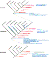
Chordate phylogeny and a synopsis of telencephalon evolution. A nested cladogram depicts the evolutionary relationships of the chordates and their relatives within the Bilateria, the group of all bilaterally symmetric animals. Red arrows and text identify common ancestors and approximate times of divergence in millions of years before the present (mya). Blue arrows and text identify the emergence of key innovations related to the evolution of the telencephalon and of the neocortex in particular. Blue text along the stems of the vertebrate groups identifies telencephalic morphological innovations of those lineages. Note that our placement of neocortical cell type origins with stem amniotes is a conservative one, based on clades for which extensive molecular data exist and are concordant with connections. See main text for discussion and references.
Figure 4.
Figure 4.
Evolution of telencephalic sensory centers in amniotes. Left: reconstruction of telencephalon organization in a stem mammal based on a comparative analysis of neocortex in extant mammals, combined with information about brain proportions from early mammalian fossil skull endocasts (adapted from Kaas [95]). This mammal is inferred to have possessed a highly developed olfactory bulb and olfactory cortex, with a compact neocortex located dorsally. This small neocortex nonetheless contained a range of neocortical areas thought to be shared in all extant mammals, a subset of which are identified here (see [95] for further discussion). The primary visual area (V1) receives lemniscal visual input (which relays through the lateral geniculate nucleus of the dorsal thalamus), whereas the middle temporal visual area (MT) receives input from a separate, parallel visual pathway that relays through the optic tectum and then the thalamic lateral posterior nucleus. All mammals additionally share a primary auditory area (A1), a primary somatosensory area (S1) and an adjoining second somatosensory area (S2). Note that this nomenclature of cortical areas does not apply to birds and non-avian reptiles, which lack a neocortex. Middle: the sizes of neocortical sensory areas do not scale linearly with the overall surface area of the neocortical sheet. That is, mammals with a highly expanded neocortex, such as humans, have a larger proportion of non-primary-sensory and higher order association cortex. Differential allocation of cortical surface area is apparent, for example, in the large human frontal cortex rostral to S1. Placement of cortical areas based on [193], MT placement in opossum and human based on [197] and [44], respectively. Right: the core sensory pathways to the mammalian pallium are conserved also in birds and non-avian reptiles, where they target spatially discrete pallial domains. The lemniscal visual channel targets the dorsal cortex (DC) in turtles and the avian Wulst (W), whereas the trans-tectal visual channel targets defined nuclei deep within the dorsal ventricular ridge (DVR) in each species [106]. Primary somatosensory information targets the DC and the Wulst rostral to the lemniscal visual targets. Primary auditory information reaches the DVR in all known sauropsids. Birds possess an additional sensory nucleus in the rostral DVR, the nucleus basorostralis, which receives trigeminal somatosensory information via a direct projection from the hindbrain [6]. This nucleus expresses molecular markers of neocortical input neurons and is conserved in alligators, but possible homologies with mammalian features remain elusive [112]. Turtle and chicken schematics adapted from [114] and [120], respectively. Cb, cerebellum; Di, diencephalon; Hb, hindbrain; Mb, midbrain; OB, olfactory bulb.
Figure 5.
Figure 5.
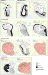
The telencephala of anamniote vertebrates. Schematics depict telencephalon anatomy from representatives of the anamniote vertebrates. Solid black territories represent regions of particularly high cell density, often along the surface of the lateral ventricle (V), whereas black dots provide a qualitative representation of cellular density and distribution. These illustrations are intended to provide a broad overview of telencephalon morphology in anamniotes and only the subset of neuroanatomical zones referred to in the main text are identified here. In all cases, pallial-subpallial boundaries should be regarded as approximations. See source materials for further details and discussion. Bullfrog adapted from [198] (DP, dorsal pallium; LP, lateral pallium; MP, medial pallium). Lungfish adapted from [24] and coelacanth from [199]. Ray-finned fishes adapted from [130] (DL, dorsolateral area; DM, dorsomedial area). Chondrichthyans adapted from [153]. Lamprey adapted from [175] (ICL, inner cellular layer; OCL, outer cellular layer; ML, molecular layer) and hagfish from [167].

Similar articles

Cited by

References

    1. Gee H (2018). Across the Bridge: Understanding the Origin of the Vertebrates (Chicago: The University of Chicago Press; ).
    1. Erwin DH, and Valentine JW (2013). The Cambrian Explosion: The Construction of Animal Biodiversity (Greenwood Village, Colorado: Roberts and Company; ).
    1. Gans C, and Northcutt RG (1983). Neural crest and the origin of vertebrates: a new head. Science 220, 268–273. - PubMed
    1. Nieuwenhuys R, Donkelaar H.J.t., and Nicholson C (1998). The Central Nervous System of Vertebrates (New York: Springer; ).
    1. Butler AB, and Hodos W (2005). Comparative Vertebrate Neuroanatomy: Evolution and Adaptation, 2nd Edition. (Hoboken, NJ: Wiley-Interscience; ).

Publication types

MeSH terms

LinkOut - more resources