. 2019 Aug 16;125(5):552-566.
doi: 10.1161/CIRCRESAHA.119.315491. Epub 2019 Jul 10.

Generation of Quiescent Cardiac Fibroblasts From Human Induced Pluripotent Stem Cells for In Vitro Modeling of Cardiac Fibrosis

Affiliations

Generation of Quiescent Cardiac Fibroblasts From Human Induced Pluripotent Stem Cells for In Vitro Modeling of Cardiac Fibrosis

Hao Zhang et al. Circ Res. .

Abstract

Rationale: Activated fibroblasts are the major cell type that secretes excessive extracellular matrix in response to injury, contributing to pathological fibrosis and leading to organ failure. Effective anti-fibrotic therapeutic solutions, however, are not available due to the poorly defined characteristics and unavailability of tissue-specific fibroblasts. Recent advances in single-cell RNA-sequencing fill such gaps of knowledge by enabling delineation of the developmental trajectories and identification of regulatory pathways of tissue-specific fibroblasts among different organs.

Objective: This study aims to define the transcriptome profiles of tissue-specific fibroblasts using recently reported mouse single-cell RNA-sequencing atlas and to develop a robust chemically defined protocol to derive cardiac fibroblasts (CFs) from human induced pluripotent stem cells for in vitro modeling of cardiac fibrosis and drug screening.

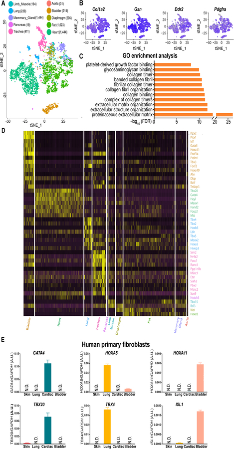
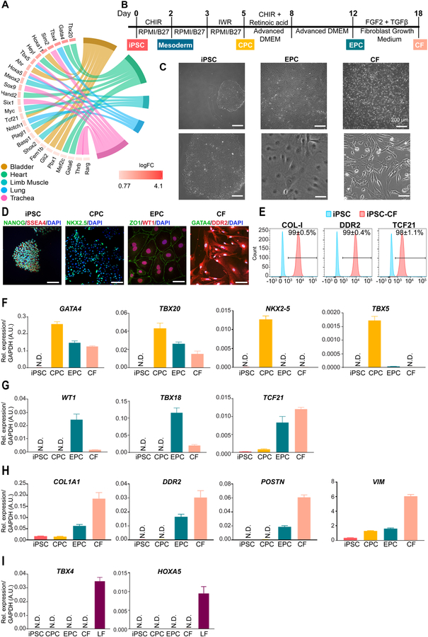
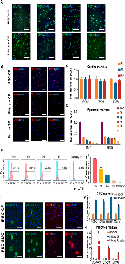
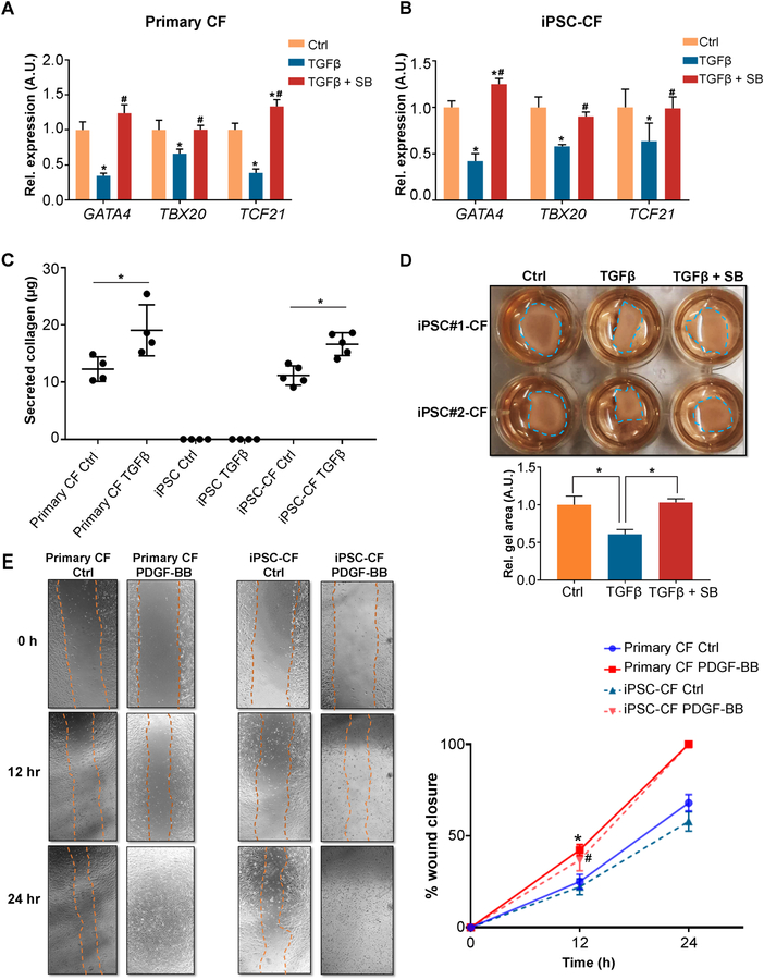
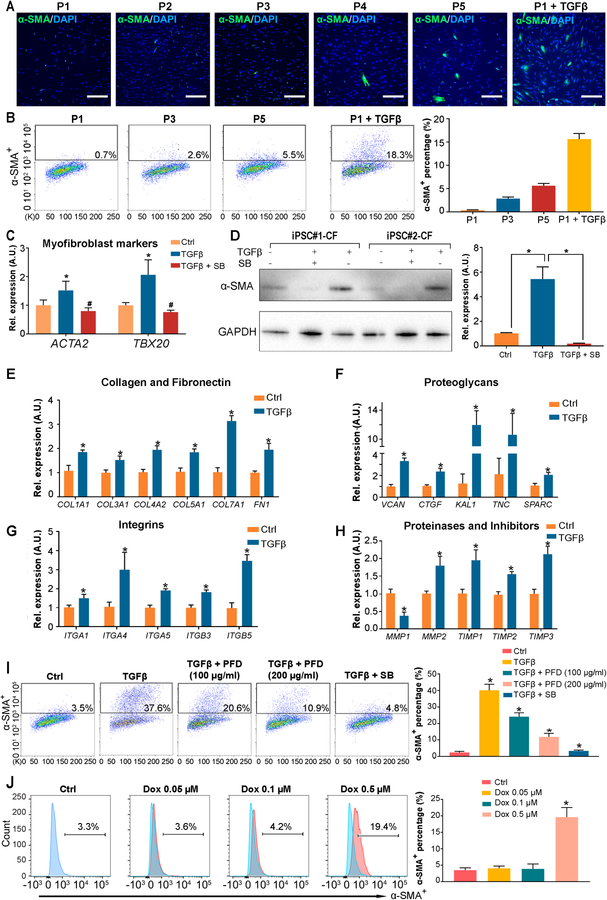
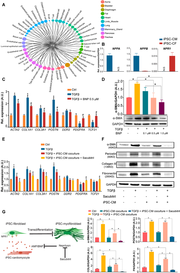
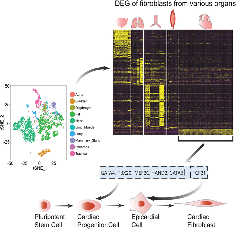
Methods and results: By analyzing the single-cell transcriptome profiles of fibroblasts from 10 selected mouse tissues, we identified distinct tissue-specific signature genes, including transcription factors that define the identities of fibroblasts in the heart, lungs, trachea, and bladder. We also determined that CFs in large are of the epicardial lineage. We thus developed a robust chemically defined protocol that generates CFs from human induced pluripotent stem cells. Functional studies confirmed that iPSC-derived CFs preserved a quiescent phenotype and highly resembled primary CFs at the transcriptional, cellular, and functional levels. We demonstrated that this cell-based platform is sensitive to both pro- and anti-fibrosis drugs. Finally, we showed that crosstalk between human induced pluripotent stem cell-derived cardiomyocytes and CFs via the atrial/brain natriuretic peptide-natriuretic peptide receptor-1 pathway is implicated in suppressing fibrogenesis.

Conclusions: This study uncovers unique gene signatures that define tissue-specific identities of fibroblasts. The bona fide quiescent CFs derived from human induced pluripotent stem cells can serve as a faithful in vitro platform to better understand the underlying mechanisms of cardiac fibrosis and to screen anti-fibrotic drugs.

Keywords: fibroblasts; fibrosis; induced pluripotent stem cells; transcriptome.


Conflict of interest statement

DECLARATION OF INTERESTS

The authors declare no competing interests.

Figures

Figure 1.
Figure 1.. Mouse single-cell transcriptome reveals tissue-specific gene markers for fibroblasts are conserved in humans.
A, A t-SNE (t-distributed stochastic neighbor embedding) plot showing the distribution patterns of 4, 685 fibroblasts derived from 10 tissues of healthy adult mice. The numbers of tissue-specific fibroblasts used for transcriptome analysis are listed in the brackets next to the individual tissue types. B, Representative t-SNE plots showing tissue-specific fibroblast subpopulations express genes reported to be detected in fibroblasts with high abundance. C, Gene ontology (GO) enrichment analysis reveals that all the cell clusters in (A) possess fibroblast-specific biological functions. D, A heatmap comparing the most specifically expressed (25% fibroblasts expressed, logFC≥1.5, and FDR adjusted P-value <1%; Wilcoxon rank sum test) transcription factors in 10 tissue-specific mouse fibroblast subpopulations. E, Cross-species validation of cardiac (GATA4 and TBX20), lung (HOXA5 and TBX4), and bladder (HOXA11 and ISL1) specific genes in human primary cardiac, lung, and bladder fibroblasts. Primary skin fibroblasts are used as a negative control. N.D., not detected. Data are presented as mean ± SEM.
Figure 2.
Figure 2.. Generation human induced pluripotent stem cell-derived cardiac fibroblasts.
A, Developmental pathway enrichment analysis (FDR adjusted P-value <1%; Hypergeometric test) showing tissue-specific transcription factors (≥25% fibroblasts, logFC≥log2 and FDR adjusted P-value <1%; Wilcoxon rank sum test) in fibroblasts of different tissue origins. B, A schematic diagram showing the protocol for small molecule-directed differentiation from human iPSCs to iPSC-CFs. iPSC, induced pluripotent stem cell; CPC, cardiac progenitor cell; EPC, epicardial cell; CF, cardiac fibroblast; FGF2, fibroblast growth factor-2; TGF-β, transforming growth factor-β. C, Representative bright-field images showing stage-specific cell morphology changes during the differentiation of human iPSC-CFs. Scale bars, 200 μm. D, Representative immunofluorescent images showing stage-specific cells express pluripotency genes (NANOG and SSEA4), cardiac mesodermal gene (NKX2–5), epicardial genes (ZO1 and WT1), and CF genes (GATA4 and DDR2) during differentiation. Scale bars, 100 μm. E, Representative flow cytometric histograms showing protein levels of COL-I, DDR2, and TCF21 in human iPSC-CFs on day 18 of differentiation (red) versus undifferentiated iPSCs (blue). F, The expression levels of cardiac-specific, early development-related genes (GATA4, TBX20, NKX2–5, and TBX5) at individual differentiation stages. N.D., not detected. G, The expression levels of epicardial markers (WT1, TBX18, and TCF21) at individual differentiation stages of iPSC-CFs. H, The expression levels of genes that are highly expressed in fibroblasts (COL1A1, DDR2, POSTN, and VIM) in stage-specific cells during iPSC-CF differentiation. I, The expression levels of lung-specific genes (TBX4 and HOXA5) in stage-specific cells during iPSC-CF differentiation. Primary LFs (lung fibroblasts) are used as a positive control. Data in (E-I) were generated based on three independent differentiations. Data are presented as mean ± SEM.
Figure 3.
Figure 3.. Human iPSC-derived cardiac fibroblasts are highly pure and preserve their cardiac identity during passaging.
A, Representative immunofluorescent images showing human iPSC-CFs are positive for gene makers (COL-I, DDR2, VIM, and POSTN) that are highly expressed in primary CFs. Scale bars, 100 μm. iPSC, induced pluripotent stem cell; CF, cardiac fibroblast. B, Representative immunofluorescent images showing the expression levels of cardiac-specific genes (GATA4, TBX20, and TCF21) in human iPSC-CFs, primary cardiac and skin fibroblasts. SF, skin fibroblast. Scale bars, 100 μm. C, The expression levels of cardiac-specific genes (GATA4, TBX20, and TCF21) in human iPSC-CFs at different passages. D, The expression levels of epicardial marker genes (WT1 and TBX18) in human iPSC-EPCs and iPSC-CFs at different passages. EPC, epicardial cell. E, Representative flow cytometric histograms showing the percentage of WT1+ population in human iPSC-EPCs and iPSC-CFs at different passages. Primary CFs are used as a control. The quantitative data are shown on the right. F, Representative immunofluorescent images showing negligible expression of SMC (smooth muscle cell)-specific genes (ACTA2, CNN1, SMTN, and TAGLN) in iPSC-CFs. The same iPSC lines were used to derive SMCs of the epicardial lineage, which are used as a positive control. Scale bars, 100 μm. G, The expression levels of SMC-specific genes (ACTA2, CNN1, MYH11, and TAGLN) in human iPSC-CFs versus iPSC-SMCs of the epicardial lineage. H, The expression levels of pericyte-specific genes (PDGFRB, CSPG4, and MCAM) in human iPSC-CFs, primary cardiac fibroblasts, and brain pericytes. Data in (C, D, E, G, and H) are generated from three independent iPSC lines, and are presented as mean ± SEM. *P<0.05 by one-way ANOVA followed by Bonferroni multiple comparisons.
Figure 4.
Figure 4.. Human iPSC-derived cardiac fibroblasts demonstrate similar biological characteristics to primary cardiac fibroblasts.
A, Expression levels of cardiac-specific genes (GATA4, TBX20, and TCF21) in human primary CFs in the absence or presence of TGF-β ± a TGF-β inhibitor SB431542 (SB). CF, cardiac fibroblast. *P<0.05 vs. vehicle control, #P<0.05 vs. TGF-β group by one-way ANOVA followed by Bonferroni multiple comparisons. B, Expression levels of cardiac-specific genes (GATA4, TBX20, and TCF21) in human iPSC-CFs in the absence or presence of TGF-β ± SB. iPSC, induced pluripotent stem cell. *P<0.05 vs. vehicle control, #P<0.05 vs. TGF-β group by one-way ANOVA followed by Bonferroni multiple comparisons. C, Secreted collagen contents (measured by Sirius red) are increased in both human iPSC-CFs and primary CFs after TGF-β treatment. Human iPSCs were used as a negative control. *P<0.05 by one-way ANOVA followed by Bonferroni multiple comparisons. D, Representative collagen gel contraction responses in two independent human iPSC-CFs in the absence or presence of TGF-β ± SB. The residual surface areas of collagen gel in each group were used for quantification (shown below the representative image), and normalized to that of the vehicle control. The size of the collagen gel area is inversely proportional to the contractility. *P<0.05 by one-way ANOVA followed by Bonferroni multiple comparisons. E, Representative images showing human iPSC-derived and primary CF migration in the absence or presence of PDGF-BB (25 ng/ml) at different time points over the 24-hour observation frame using a wound-healing assay. Quantitative data showing similar migration rate (wound closure area) between human iPSC-CFs and primary CFs under the same conditions. *P<0.05 indicates PDGF-BB treatment vs. vehicle control in primary CF, #P<0.05 indicates PDGF-BB treatment vs. vehicle control in iPSC-CF, by one-way ANOVA followed by Bonferroni multiple comparisons. All data were generated from three or four independent iPSC lines, and are presented as mean ± SEM.
Figure 5.
Figure 5.. Human iPSC-derived cardiac fibroblasts are quiescent fibroblasts and therefore provide an ideal in vitro platform to model fibrosis.
A-B, Representative immunofluorescent (A) and flow cytometry (B) images showing the degree of spontaneous transdifferentiation of iPSC-CFs to myofibroblasts (α-SMA+, smooth muscle α-actin) during passages 1–5. TGF-β treated CFs were used as a positive control. The percentage of SMA+ myofibroblasts in each passage of iPSC-CFs are calculated based on the flow cytometry (B, right). iPSC, induced pluripotent stem cell; CF, cardiac fibroblast. Scale bars, 100 μm. C, The expression levels of myofibroblast markers (ACTA2 and POSTN) in human iPSC-CFs cultured in the absence or presence of TGF-β ± a TGF-β inhibitor SB431542 (SB). *P<0.05 vs. vehicle control, #P<0.05 vs. TGF-β group by one-way ANOVA followed by Bonferroni multiple comparisons. D, A representative immunoblot showing the α-SMA levels in CFs derived from two independent iPSC lines in the absence or presence of TGF-β ± SB. The corresponding densitometric quantification data are shown next to the immunoblot. *P<0.05 by one-way ANOVA followed by Bonferroni multiple comparisons. E-H, The expression levels of genes coding extracellular matrix proteins (collagen, fibronectin, proteoglycans, integrins, and matrix proteinases and inhibitors) in human iPSC-CFs cultured in the absence or presence of TGF-β. *P<0.05 vs. vehicle control by one-way ANOVA followed by Bonferroni multiple comparisons. I, Representative flow cytometry images and quantitative data showing pirfenidone (a commercial anti-fibrosis drug) or SB significantly suppresses TGF-β induced propagation of the α-SMA+ population in iPSC-CFs. PFD, pirfenidone. *P<0.05 vs. TGF-β group by one-way ANOVA followed by Bonferroni multiple comparisons. J, Representative flow cytometric histograms and quantitative data showing a dose-dependent induction of the α-SMA+ population after human iPSC-CFs were treated with doxorubicin. *P<0.05 vs. ctrl group by one-way ANOVA followed by Bonferroni multiple comparisons. Data in (B-J) were generated from three independent iPSC lines, and are presented as mean ± SEM.
Figure 6.
Figure 6.. Human iPSC-derived cardiomyocytes attenuate TGF-β-induced fibrotic responses in iPSC-derived cardiac fibroblasts through a paracrine mechanism.
A, Computational analysis showing broad intercellular communications between fibroblasts and a diverse array of cell types through different complementary ligand-receptor signaling pathways. The threshold for defining a ligand/receptor expressed in a cluster is ≥25% with gene expression > 0. FB, fibroblast. B, The basal expression levels of ligands NPPA (natriuretic peptide type A) and NPPB and their cognate receptor NPR1 (natriuretic peptide receptor 1) in human iPSC-CMs and iPSC-CFs. iPSC, induced pluripotent stem cell; CM, cardiomyocyte; CF, cardiac fibroblast. C, Expression levels of CF and myofibroblast markers in human iPSC-CFs in the absence or presence of TGF-β ± BNP. *P<0.05 vs. vehicle control, #P<0.05 vs. TGF-β group by one-way ANOVA followed by Bonferroni multiple comparisons. D, A representative immunoblot showing α-SMA (smooth muscle α-actin) levels in human iPSC-CFs treated with TGF-β and escalating concentrations of BNP. Densitometric quantifications are shown above the immunoblot. *P<0.05 by one-way ANOVA followed by Bonferroni multiple comparisons. E, Expression levels of CF and myofibroblast markers in human iPSC-CFs after they were indirectly co-cultured with or without iPSC-CMs in the absence or presence of TGF-β ± sacubitril. Sacubitril is an inhibitor for ANP (atrial natriuretic peptide) and BNP-degrading enzyme. *P<0.05 vs. vehicle control, #P<0.05 vs. TGF-β group, $P<0.05 vs. iPSC-CM co-culture group by one-way ANOVA followed by Bonferroni multiple comparisons. F, Representative immunoblots showing the levels of α-SMA, periostin (POSTN), collagen III (COLIII), and fibronectin (FN) in human iPSC-CFs after they were indirectly co-cultured with or without iPSC-CMs in the absence or presence of TGF-β ± sacubitril. Densitometric quantifications are shown below the immunoblots. *P<0.05 by one-way ANOVA followed by Bonferroni multiple comparisons. G, Schematic of the proposed mechanisms for the role of cardiomyocytes in suppressing TGF-β-induced CF transdifferentiation through the ANP/BNP-NPR1 signaling pathway. Sacubitril, a pharmacological drug, can further enhance the anti-fibrotic effect by suppressing the activity of ANP- and BNP-degrading enzyme, neprilysin. Data in (B-F) were generated based on three independent differentiations, and are presented as mean ± SEM.
Figure 7.
Figure 7.. A logical flow diagram demonstrates the process of developing the human iPSC-derived cardiac fibroblast differentiation protocol.
The differentiation protocol for human iPSC -CFs developed in this study is based on the recognition of tissue-specific marker genes expressing in fibroblast subpopulations, as revealed by the mouse single-cell transcriptomic data. Further DEG (differentially expressed genes) analysis highlights the critical roles of tissue-specific transcription factors in regulating the development trajectories of fibroblast subpopulations, and also suggests that CFs may be derived from the epicardial lineage. As such, human iPSC-CFs were successfully generated after intermediate cell types of cardiac progenitor cells and epicardial cells being sequentially differentiated. iPSC, induced pluripotent stem cell; CF, cardiac fibroblast.

Comment in

References

    1. Tomasek JJ, Gabbiani G, Hinz B, Chaponnier C and Brown RA. Myofibroblasts and mechano-regulation of connective tissue remodelling. Nat Rev Mol Cell Biol. 2002;3:349–63. - PubMed
    1. Zeisberg M and Kalluri R. Cellular mechanisms of tissue fibrosis. 1. Common and organ-specific mechanisms associated with tissue fibrosis. Am J Physiol Cell Physiol. 2013;304:C216–25. - PMC - PubMed
    1. Paik DT, Tian L, Lee J, Sayed N, Chen IY, Rhee S, Rhee JW, Kim Y, Wirka RC, Buikema JW, Wu SM, Red-Horse K, Quertermous T and Wu JC. Large-scale single-cell RNA-seq reveals molecular signatures of heterogeneous populations of human induced pluripotent stem cell-derived endothelial cells. Circ Res. 2018;123:443–450. - PMC - PubMed
    1. Shi Y, Inoue H, Wu JC and Yamanaka S. Induced pluripotent stem cell technology: a decade of progress. Nat Rev Drug Discov. 2017;16:115–130. - PMC - PubMed
    1. The Tabula Muris Consortium. Single-cell transcriptomics of 20 mouse organs creates a Tabula Muris. Nature. 2018;562:367–372. - PMC - PubMed

Publication types

Substances