. 2019 Jul 9;12(1):104.
doi: 10.1186/s12920-019-0552-1.

Retroposed copies of RET gene: a somatically acquired event in medullary thyroid carcinoma

Affiliations

Retroposed copies of RET gene: a somatically acquired event in medullary thyroid carcinoma

Larissa V Bim et al. BMC Med Genomics. .

Abstract

Background: Different pathogenic germline mutations in the RET oncogene are identified in MEN 2, a hereditary syndrome characterized by medullary thyroid carcinoma (MTC) and other endocrine tumors. Although genetic predisposition is recognized, not all RET mutation carriers will develop the disease during their lifetime or, likewise, RET mutation carriers belonging to the same family may present clinical heterogeneity. It has been suggested that a single germline mutation might not be sufficient for development of MEN 2-associated tumors and a somatic bi-allelic alteration might be required. Here we investigated the presence of somatic second hit mutation in the RET gene in MTC.

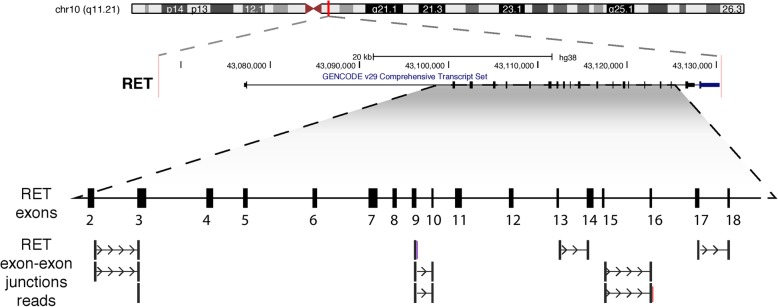
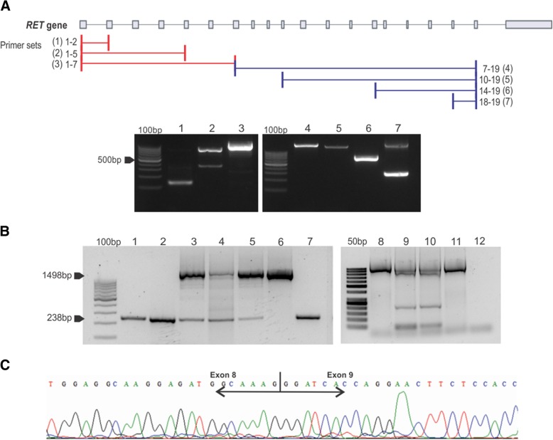
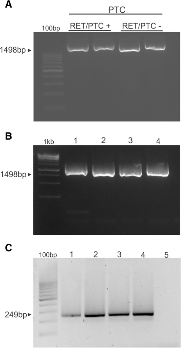
Methods: We integrated Multiplex Ligation-dependent Probe Amplification (MLPA) and whole exome sequencing (WES) to search for copy number alteration (CNA) in the RET gene in MTC samples and medullary thyroid cell lines (TT and MZ-CR-1). We next found reads spanning exon-exon boundaries on RET, an indicative of retrocopy. We subsequently searched for RET retrocopies in the human reference genome (GRCh37) and in the 1000 Genomes Project data, by looking for reads reporting joined exons in the RET locus or distinct genomic regions. To determine RET retrocopy specificity and recurrence, DNA isolated from sporadic and MEN 2-associated MTC (n = 37), peripheral blood (n = 3) and papillary thyroid carcinomas with RET fusion (n = 10) samples were tested using PCR-sequencing methodology.

Results: Through MLPA we have found evidence of CNA in the RET gene in MTC samples and MTC cell lines. WES analysis reinforced the presence of the CNA and hinted for a retroposed copy of RET not found in the human reference genome and 1.000 Genomes Project. Extended analysis confirmed the presence of a somatic MTC-related retrocopy of RET in both sporadic and hereditary tumors. We further unveiled a recurrent (28%) novel point mutation (p.G548 V) found exclusively in the retrocopy of RET. The mutation was also found in cDNA of mutated samples, suggesting it might be functional.

Conclusion: We here report a somatic specific RET retroposed copy in MTC samples and cell lines. Our results support the idea that generation of retrocopies in somatic cells is likely to contribute to MTC genesis and progression.

Keywords: G548V; MEN 2; MTC; Medullary thyroid carcinoma; RET; Retrocopy; Second hit.


Conflict of interest statement

The authors reported no conflicts of interest.

Figures

Fig. 1
Fig. 1
Chart obtained by Coffalyzer analysis showing relative probe ratio values from MLPA results of RET gene analysis. The upper and lower bars represent the threshold values of normality. MLPA shows normal ratio for RET exons 1–20 in (a) normal thyroid tissue (b) peripheral blood and (c) C-cell hyperplasia, corroborating normal copy numer. Bullets located upper the duplication cut-off line (upper bar) in the ratio chart were identified in (d) Medullary Thyroid Carcinoma (MTC) and MTC- derived cell lines (E) MZ-CRC-1 cell line and (f) TT cell line, which denoted somatic duplication
Fig. 2
Fig. 2
Genomic representation of whole exome sequencing data showing reads spanning exon-exon boudaries. We found reads spanning 10 exon-exon boundaries, from exon 2 to exon 18 (considering transcripts with the highest number of exons annotated). Representation based on UCSC Genome browser plot for the RET locus
Fig. 3
Fig. 3
Detection of Somatic retrocopies. (a) Schematic figure representing the RET parental gene and the primer sets (1–7) used to amplify the RET retrocopy. The line represents the non conding sequence (introns and UTR) and the bars represent the exons. The agarose gel shows bands at the expected sizes for intronless copy for all primer sets tested, indicating the presence of a retrocopy of RET in genomic DNA isolated from MTC. As expected, the band corresponding to the intron-containing fragment (parental copy) was observed only for primer set 7 (1868pb). The additional band indentified in line 2 of primer set 2 is an inespecific PCR product (Additional file 2 :Table S2). (b) Representative PCR analysis performed in MTC and control samples using primers located at exons 7 and 9 (primer set 8, Additional file 2: Table S2) of RET gene. The upper band corresponds to the expected PCR product (1498 bp) for RET parental gene and the lower band (238 bp) corresponds to the retrocopy (intronless copy). The lower band is seen only in tumor samples (Lines 1–5) and MTC derived cell lines (lines 9 and 10). Lines 1 and 2 were FFPE derived sample and lines 3–5 are fresh frozen tissue drived samples. The lower band was not seen in peripheral blood DNA from patients (line 6), non-MTC cell line (line 11) and MTC cell line obtained from ATCC (third passage). Positive (line 7) and Negative (line 12) controls were used. (c) Sequencing analysis of the retrocopy band (238 bp) confirming exon-exon junction
Fig. 4
Fig. 4
Detection of somatic retrocopies in (a) PTC samples positive or negative for the RET/PTC rearrangent and (b) neuroblastoma (line 1), craniopharyngioma (line 2), pilocytic astrocytoma (line 3) and ependymoma (line 4). (c) Amplification of KRASP1 (processed pseudogene) in MTC (line 1) peripheral blood (line 2) normal thyroid tissue (line 3) positive control (line 4) and negative control (line 5)
Fig. 5
Fig. 5
(a) Schematic representation of exons 7 to 9 of the RET parental gene and retrocopy. Rectangles denote exons, full line denotes introns. The arrow marks the location of the exon 8 p.G548V mutation. The horizontal arrows indicate the location and orientation of primers used. (b) Prevalence of the p.G548V mutation on MTC samples, showing that 27,9% of all MTC tested samples are positive. (c) Representative electropherogram showing the presence of the mutation both in heterozygosis and homozygosis. The mutations consist of a genomic 1644G > T change. (d) Sequencing of DNA amplification from parental RET gene and its retrocopy from MZ-CRC-1 cell line showing the p.G548V mutation present only in the retrocopy

References

    1. Wells SA Jr, Pacini F, Robinson BG, Santoro M. Multiple endocrine neoplasia type 2 and familial medullary thyroid carcinoma: an update. J Clin Endocrinol Metab. 2013. - PMC - PubMed
    1. Mulligan LM, Eng C, Healey CS, Clayton D, Kwok JB, Gardner E, Ponder MA, Frilling A, Jackson CE, Lehnert H, et al. Specific mutations of the RET proto-oncogene are related to disease phenotype in MEN 2A and FMTC. Nat Genet. 1994;6:70–74. doi: 10.1038/ng0194-70. - DOI - PubMed
    1. American Thyroid Association Guidelines Task F Kloos RT, Eng C, Evans DB, Francis GL, Gagel RF, Gharib H, Moley JF, Pacini F, Ringel MD, et al: medullary thyroid cancer: management guidelines of the American Thyroid Association. Thyroid. 2009;19:565–612. doi: 10.1089/thy.2008.0403. - DOI - PubMed
    1. Pacak K, Eisenhofer G, Ilias I. Diagnosis of pheochromocytoma with special emphasis on MEN2 syndrome. Hormones (Athens) 2009;8:111–116. doi: 10.14310/horm.2002.1227. - DOI - PMC - PubMed
    1. Eng C, Clayton D, Schuffenecker I, Lenoir G, Cote G, Gagel RF, van Amstel HK, Lips CJ, Nishisho I, Takai SI, et al. The relationship between specific RET proto-oncogene mutations and disease phenotype in multiple endocrine neoplasia type 2. International RET mutation consortium analysis. JAMA : the journal of the American Medical Association. 1996;276:1575–1579. doi: 10.1001/jama.1996.03540190047028. - DOI - PubMed

Publication types

Supplementary concepts