. 2020 Apr;105(4):987-9998.
doi: 10.3324/haematol.2018.214379. Epub 2019 Jul 9.

Mesenchymal stromal cells confer chemoresistance to myeloid leukemia blasts through Side Population functionality and ABC transporter activation

Affiliations

Mesenchymal stromal cells confer chemoresistance to myeloid leukemia blasts through Side Population functionality and ABC transporter activation

Laetitia Boutin et al. Haematologica. 2020 Apr.

Abstract

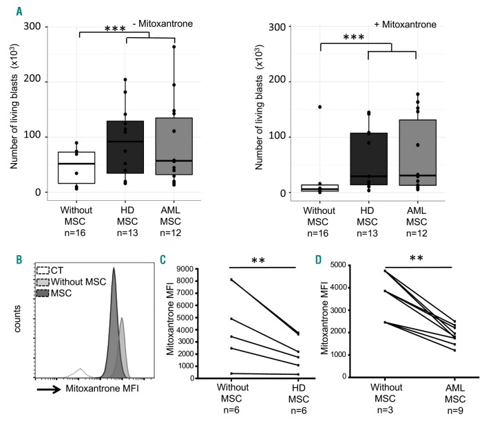
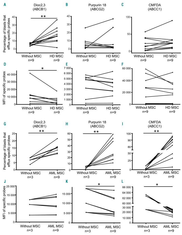
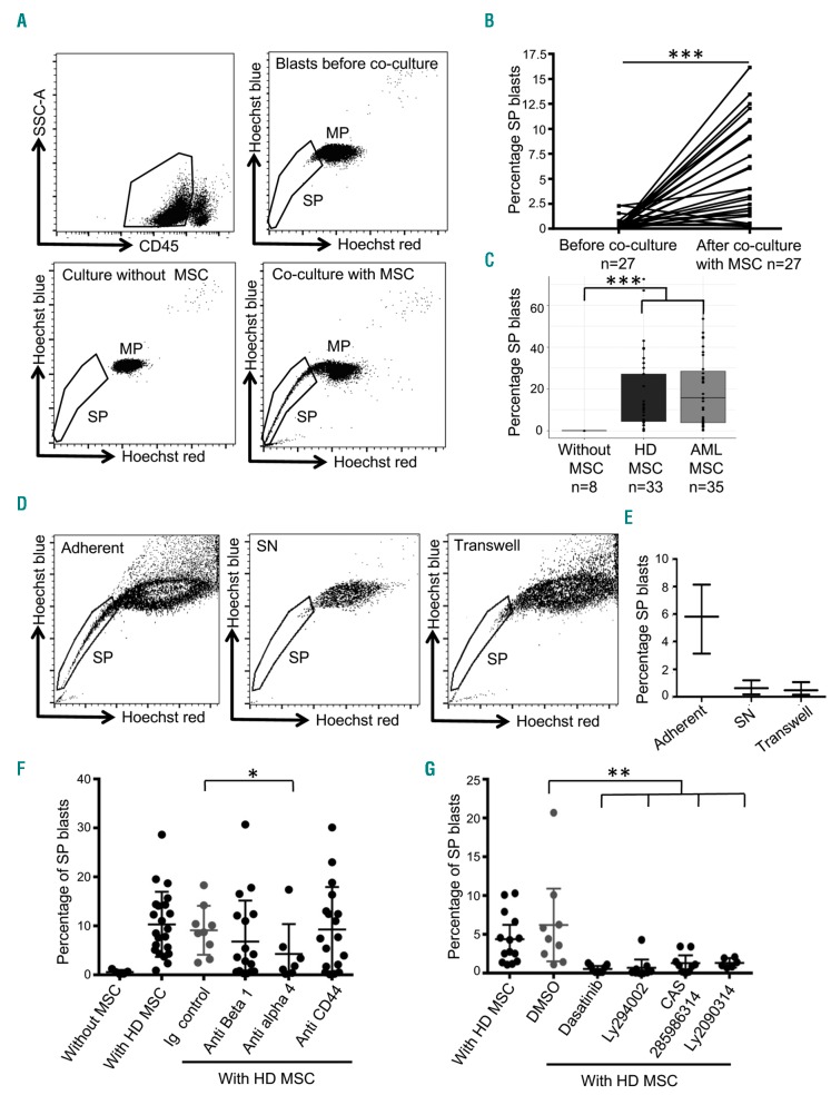
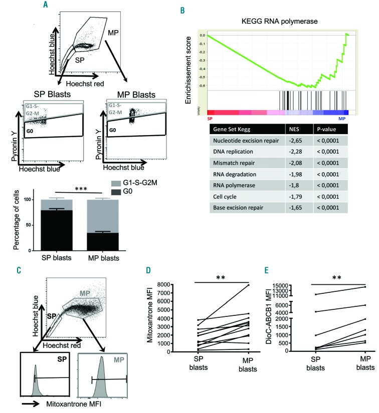
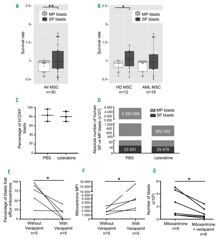
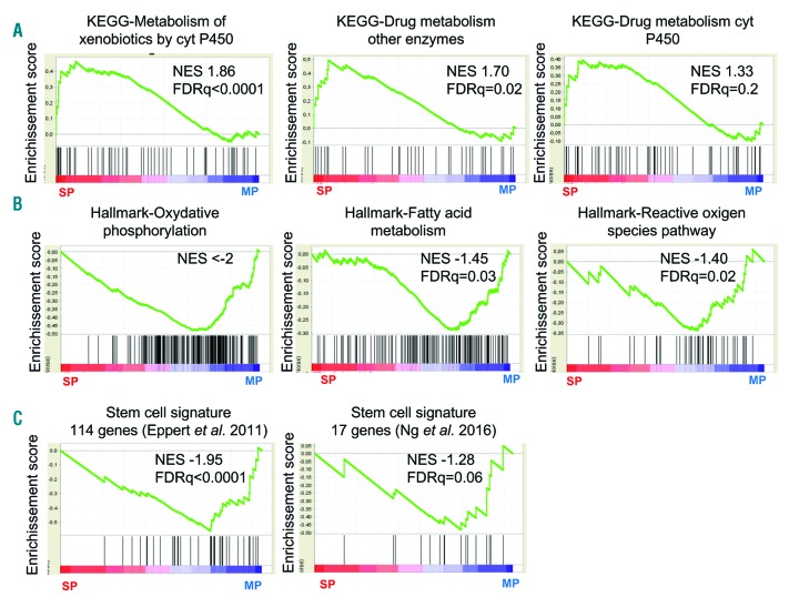
Targeting chemoresistant malignant cells is one of the current major challenges in oncology. Therefore, it is mandatory to refine the characteristics of these cells to monitor their survival and develop adapted therapies. This is of particular interest in acute myeloid leukemia (AML), for which the 5-year survival rate only reaches 30%, regardless of the prognosis. The role of the microenvironment is increasingly reported to be a key regulator for blast survival. In this context, we demonstrate that contact with mesenchymal stromal cells promotes a better survival of blasts in culture in the presence of anthracycline through the activation of ABC transporters. Stroma-dependent ABC transporter activation leads to the induction of a Side Population (SP) phenotype in a subpopulation of primary leukemia blasts through alpha (α)4 engagement. The stroma-promoting effect is reversible and is observed with stromal cells isolated from either healthy donors or leukemia patients. Blasts expressing an SP phenotype are mostly quiescent and are chemoresistant in vitro and in vivo in patient-derived xenograft mouse models. At the transcriptomic level, blasts from the SP are specifically enriched in the drug metabolism program. This detoxification signature engaged in contact with mesenchymal stromal cells represents promising ways to target stroma-induced chemoresistance of AML cells.


Figures

Figure 1.
Figure 1.
Mesenchymal stromal cells (MSC) from heathy donors (HD) or from acute myeloid leukemia (AML) patients confer a better survival to leukemia blasts, even in the presence of chemotherapy agents. (A, left) Histogram representing the number of living AML blasts after a 3-day culture without MSC [median 51,655 (56,830)] or with MSC from HD [median 91,815 (94,556)] or AML patients [median 56,896 (102,863)]. (Right) The same conditions but in the presence of 50nM of mitoxantrone. Culture conditions in the presence of the different types of MSC confer a significantly improved survival to AML blasts (left: ***P=0.0004; right: ***P=0.0003, n>10, represents the number of AML blasts/MSC donor combinations). (B) Histogram showing fluorescence intensity of AML blasts stained with mitoxantrone after a 3-day culture with or without MSC from HD (one representative experiment of the 6 performed). Control (CT) represents non-stained blasts. (C) Mitoxantrone mean fluorescence intensity (MFI) in AML blasts cultivated [median 2,191 (2,032)] or not [median 3,676 (3,546)] on HD MSC (**P=0.01, n=6, Wilcoxon test). (D) Mitoxantrone MFI in AML blasts cultivated [median 3,861 (1,147)] or not [median 1,821 (460)] on AML MSC (**P=0.01, n=3-9, Wilcoxon test). n: number. **P<0.01; ***P<0.001.
Figure 2.
Figure 2.
Co-culture between acute myeloid leukemia (AML) blasts and mesenchymal stromal cells (MSC) modulates the ABC transporter functionality on leukemia blasts in a patient-dependent manner. (A-C) Percentage of AML blasts that efflux specific probes (ABC transporter activity) after a 3-day co-culture on healthy donor (HD) MSC. ABC transporter activities were quantified using specific probes (Dioc2,3, Purpurin 18, CMFDA for ABCB1, ABCG2, ABCC1, respectively) as shown on the histograms. ABCB1 activity is significantly increased (P=0.0059, n=9, Wilcoxon test) by AML blasts after a 3-day co-culture on HD MSC. (D-F) Corresponding probe mean fluorescence intensity (MFI) in AML blasts; Dioc2,3 MFI is significantly decreased (P=0.02, n=9, Wilcoxon test) in blasts after co-culture with HD MSC. (G-I) Percentage of AML blasts that efflux specific probes after a 3-day co-culture on AML MSC. Activity of the ABCB1, ABCG2 and CMFDA transporters was significantly increased (P=0.01, P=0.02 and P=0.008, respectively; n=3 without MSC and n=9 with AML MSC, Wilcoxon test). (J-L) Corresponding MFI in AML blasts. Purpurin 18 and CMFDA MFI are significantly decreased (P=0.01 and P=0.01, respectively; n=3 without MSC and n=9 with AML MSC, Wilcoxon test) in co-culture conditions. *P<0.05; **P<0.01.
Figure 3.
Figure 3.
Circulating leukemia blasts acquire a Side Population (SP) phenotype in contact with mesenchymal stromal cells (MSC) from healthy donors (HD) or acute myeloid leukemia (AML) patients through α4 integrin interaction. (A) Cytograms illustrating the gating strategy of AML blasts for cytometry analysis (top left) and SP visualization before co-culture (top right) and after a 3-day culture without (bottom left) or with (bottom right) HD MSC. (B) Percentage of SP AML blasts before and after a 3-day co-culture on MSC (either from HD or AML patients) (P<0.001, n=27, Wilcoxon test). (C) Percentage of SP AML blasts after co-culture with HD MSC (P<10−4, n=8-33) or AML patients (P<10−4, n=8-35). (D) Cytograms showing SP phenotype observed on AML blasts from MSC adherent (left) or supernatant fractions (SN, middle) and in transwell experiments (right) after a 3-day co-culture with HD MSC. (E) Percentage of SP blasts in the three experimental conditions. (F and G) Percentage of SP AML blasts after co-culture or not with HD MSC and after inhibition of β1 (n=18) and α4 (n=7) integrins (P=0.04) or CD44 (n=18) (F) and their downstream signaling pathways (G) using dasatinib, LY294002, CAS2859863 and LY209031, inhibiting Src (P=0.002, n=9), AKT (P=0.003, n=9), STAT5 (P=0.009, n=9), and GSK3 pathways (P=0.01, n=6), respectively. *P<0.05; **P<0.01; ***P<0.001.
Figure 4.
Figure 4.
The mesenchymal stromal cells (MSC)-induced Side Population (SP) functionality in leukemia blasts is associated with quiescence and chemotherapy efflux through ABC transporter activation. (A) Cytograms and histograms showing gating strategies and percentage of SP and MP blasts in G0 and in G1-S-G2-M after a 3-day co-culture on HD MSC. Cell cycle status of SP versus Main Population (MP) blasts was analyzed adding pyronin Y during Hoechst staining. SP blasts are mostly in G0 [median 76% (16.5%)] compared to MP [median 33.45% (18.6%)] blasts which are in G1-S-G2-M (P=0.0009, n=12, Wilcoxon test). (B) Transcriptomic analysis of SP blasts compared to MP blasts. (C) Cytograms showing gating strategy to evaluate mitoxantrone efflux in SP or MP blasts. Quantification of mitoxantrone MFI (D) in SP [median 1,584 (1,845)] and in MP [median 3219 (1,706)] blasts (P=0.0052, n=12, Wilcoxon test). (E) DioC2,3 MFI evaluating ABCB1 activity in SP [median 239 (4,056)] and MP [median 1,298 (5418)] cells (P=0.023, n=7, Wilcoxon test). **P<0.01; ***P<0.001.
Figure 5.
Figure 5.
Side Population (SP) functionality of acute myeloid leukemia (AML) blasts induced by mesenchymal stromal cell (MSC) interactions is associated with chemoresistance. (A) Survival rate of SP [median 0.98 (0.46)] or Main Population (MP) [median 0.95 (0.24)] blasts after a 3-day co-culture with MSC in presence of mitoxantrone (P=0.041, n=30, Wilcoxon test). (B) Survival rate of SP or MP blasts after a 3-day co-culture with MSC from healthy donors (HD) or AML patients. (C) Percentage of hCD45+ blasts within the total CD45+ population. (D) Absolute number of human SP and MP blasts in the femur of patient-derived xenograft (PDX) mice after a 5-day treatment with either phosphate buffered saline or cytarabine (30 mg/kg) (n=3 mice per patient). (E) Percentage of blasts that efflux mitoxantrone with or without verapamil after a 3-day co-culture on AML MSC (P=0.05, n=5, Wilcoxon test) and (F) the corresponding mitoxantrone mean fluorescence intensity (MFI) in AML blasts (P=0.05, n=5, Wilcoxon test). (G) Number of living blasts co-cultivated on AML or HD MSC after 24 hours of mitoxantrone and treatment with or without verapamil (P=0.01, n=6, Wilcoxon test) *P<0.05; **P<0.01.
Figure 6.
Figure 6.
Side Population (SP) blasts exhibit a detoxification program signature. Gene set enrichment analysis with transcriptomics data of sorted SP and Main Population (MP) blasts after a 3-day co-culture on healthy donor (HD) mesenchymal stromal cells (MSC) shows an upregulation in SP cells of genes of xenobiotic metabolism and drug metabolism through the cytochrome P450 or other enzymes (A), an upregulation in MP blasts of genes of oxidative phosphorylation, fatty acid metabolism and reactive oxygen pathway (B), as well as of genes from the stem cell signatures reported by Eppert et al. and Ng et al. (C).

References

    1. Arber DA, Orazi A, Hasserjian R, et al. The 2016 revision to the World Health Organization classification of myeloid neoplasms and acute leukemia. Blood. 2016;127(20):2391–2405. - PubMed
    1. Taussig DC, Miraki-Moud F, Anjos-Afonso F, et al. Anti-CD38 antibody-mediated clearance of human repopulating cells masks the heterogeneity of leukemia-initiating cells. Blood. 2008;112(3):568–575. - PubMed
    1. van Rhenen A, Feller N, Kelder A, et al. High stem cell frequency in acute myeloid leukemia at diagnosis predicts high minimal residual disease and poor survival. Clin Cancer Res. 2005;11(18):6520–6527. - PubMed
    1. Vergez F, Green AS, Tamburini J, et al. High levels of CD34+CD38low/−CD123+ blasts are predictive of an adverse outcome in acute myeloid leukemia: a Groupe Ouest-Est des Leucémies Aiguës et Maladies du Sang (GOELAMS) study. Haematologica. 2011;96(12):1792–1798. - PMC - PubMed
    1. Jordan CT, Upchurch D, Szilvassy SJ, et al. The interleukin-3 receptor alpha chain is a unique marker for human acute myelogenous leukemia stem cells. Leukemia. 2000;14(10):1777–1784. - PubMed

Publication types

Substances